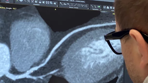
SCCT aims to provide common language for CCTA use with updated guidance
During the past decade, coronary computed tomography angiography (CCTA) has become increasingly important in clinical care and research. Cardiac CT's growth has resulted in a growing number of tools for comprehensive coronary analyses used both in research and clinical care. However, the field currently lacks standards for the quantitative image interpretation of CCTA and the reporting of results and. Clinicians and researchers need a common language so that they can communicate their findings with one another.
In a step forward to better define standards and common clinical nomenclatures, the Society of Cardiovascular Computed Tomography (SCCT) has developed an expert consensus document to help standardize quantitative assessments by CCTA.[1]
“This document provides for the first time a standardization on nomenclature for quantitative parameters derived from CCTA,” Hector M. Garcia-Garcia, MD, PhD, writing group co-chair and corresponding author, said in a statement. “It marks a turning point in reporting of CCTA as it focuses on actual quantification of atherosclerosis.”
According to the writing group, co-led by SCCT Past President Koen Nieman, MD, PhD, well-described semi-quantitative scores and classifications are primarily based on visual interpretation or through categories of obstructive severity rather than exact percentage stenosis measurements. These include Agatston scores, segment involvement scores and assessments of stenosis and plaque within the Coronary Artery Disease Reporting and Data System (CAD-RADS) in addition to disease classification scores like CT SYNTAX for coronary revascularization and CT-RECTOR Score for chronic total occlusions (CTO).
For the clinical management of patients, semiquantitative measures of coronary artery disease (CAD) are often sufficient. Existing guidelines recommend reporting of coronary stenoses by categories of obstructive severity rather than by using exact percentages.
The writing group said despite these available measurements and guidelines, the field currently lacks standards for the quantitative image interpretation of and reporting of CCTA results. The scientific document was created to fill that gap and create a common language for clinicians and researchers to communicate findings for coronary analyses.
“CCTA has been recognized as a promising noninvasive tool to monitor CAD progression and assess the effects of medical therapy or mechanical interventions in clinical trials,” the group wrote. “Specific and clear definitions as well as standardization of methodology are essential for CCTA to mature as an accurate and reproducible endpoint in clinical research. The reader should view the expert consensus document as the best attempt of the SCCT to inform and guide clinical/research practice in this area where rigorous evidence may not yet be available or the evidence to date is not widely accepted."
Additional parameters covered within the document include:
• Image acquisition, quality and analysis
• Artifacts and image degradation
• Vessel and lumen segmentation
• Deep learning-based vessel segmentation
• Serial imaging studies
• Selection of CCTA-derived study endpoints
• Future directions in CCTA technology
AI use in cardiac CT
The document highlights how deep-learning algorithms are changing CCTA assessments by enabling AI to perform much more precise measurements, rather than semiquantitative measures currently used by readers. AI of this type is already commercialized for identification of different plaque types, quantification of calcium score, and to perform lumen segmentation.
"Fully automated machine-learning algorithms, and especially deep learning segmentation methods, promise to provide a faster method for plaque analysis. In the future, fully user-free segmentation of medical images may be possible, but the application of deep-learning for CCTA has many obstacles that still must be surmounted," the authors wrote.
The biggest obstacle so far appears to be finding large enough amounts of curated image data to train AI models for image segmentation. These models also need to be subjected to careful validation using the corresponding gold standards, such as invasive coronary angiography for stenosis evaluation or IVUS for plaque quantification.
Cardiac CT experts have said AI will likely make all measurements in imaging much more precise in the coming years. Vendors offering AI for soft plaque assessment say their software is precise enough to see small changes, which can be used for screening patients, monitoring disease progression and to can better assess the impact of prevent drugs and a reversal of coronary plaque.

