
FAPI PET shows promise to make a big impact in cardiovascular imaging
A novel radiotracer originally developed to improve cancer detection using positron emission tomography (PET) is showing promise for new cardiovascular nuclear imaging indications, including the identification of pulmonary arterial hypertension (PAH) and left ventricular dysfunction following myocardial infarction.
PET tracer research focused on fibroblast activation protein inhibitors (FAPI) is quickly starting to gain momentum. It is considered one of the most promising new targets for oncology nuclear imaging because the fibroblast activation protein (FAP) is over expressed in cancer-associated fibroblasts of several types of tumors. The tracer also offers numerous advantages over the current standard of fluorine-18 (F-18) FDG imaging. These include rapid tumor uptake and a very low accumulation in normal tissues, combined with rapid clearance from the circulation. This enables high-contrast FAPI PET/CT imaging within 10 minutes after tracer administration. Another key benefit is the fact that FAPI does not require a special diet or fasting prior to imaging.
FAPI also has applications in other diseases where infection, inflammation and pro-fibrotic activity play a role, which includes numerous cardiovascular presentations.
The Prognostic Value of FAPI PET in PAH
A new study published in JACC: Cardiovascular Imaging highlighted the use of F-18 FAPI accumulation in the right ventrical (RV) and pulmonary arteries (PAs) of patients with PAH.[1] The study of 46 patients followed out to 17 months and found that uptake in the RV free wall may be a promising noninvasive prognostic indicator for PAH.
The patients were stratified into the low-, intermediate- and high-risk groups and they were observed for the primary endpoint of worsening PAH events. The F-18 FAPI uptake was quantified as target-to-background ratio (TBR) for both RV and PAs.
Significantly higher TBR in the RV and PAs were observed in clinical worsening vs non–clinical worsening patients. The study found 22 patients who experienced clinical worsening events. The authors said a multivariable Cox regression analysis identified the TBR of RV free wall (TBRRVFW) as an independent predictor of clinical worsening.
FAPI imaging to detect left ventricular dysfunction
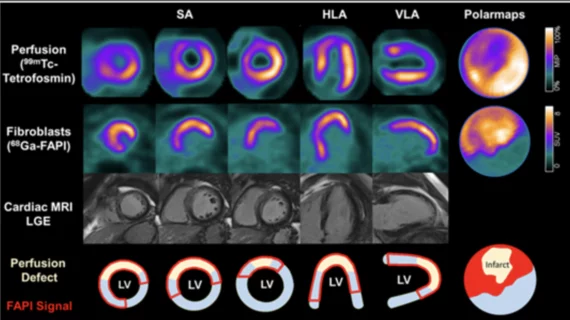
The Society of Nuclear Medicine and Molecular Imaging (SNMMI) selection of the 2022 Image of the Year at its annual meeting was from a cardiac FAPI study.
It was part of a German study for the new PET imaging agent gallium-68 (Ga-68) FAPI-46. Researchers found this agent, when correlated with cardiac MR images, offers a new perspective of what happens to a patient’s heart following an acute myocardial infarction (AMI).[2]
“FAPI is a very exciting radiotracer that holds great potential for the future of nuclear medicine and molecular imaging,” SNMMI 2022 Scientific Program chair Heather Jacene, MD, said in a statement at the time. “As shown in the Image of the Year, Ga-68 FAPI-46 PET/CT clearly indicates pro-fibrotic activity after acute myocardial infarction. The predictive value of FAPI-PET for the magnitude of subsequent decline in global cardiac function, if further validated, may be used in the future to help select the most suitable patients for anti-fibrotic therapies which are currently under development. This could ultimately have a powerful impact on cardiovascular medicine.”
A preclinical study presented at the American Heart Association 2023 meeting found Ga-68 FAPI-04 PET is a suitable tracer to image extracellular matrix remodeling and fibrosis during early cardiac injury from cancer chemotherapy anthracycline-induced cardiotoxicity.[3]
The American Society of Nuclear Cardiology (ASNC) has made a major push in recent years for a wider adoption of cardiac PET. The society's experts argue that PET not only offers better diagnostic information for cardiac perfusion over SPECT, the current standard of care, but also will allow numerous new applications for cardiovascular imaging beyond perfusion.

Dave Fornell has covered healthcare for more than 17 years, with a focus in cardiology and radiology. Fornell is a 5-time winner of a Jesse H. Neal Award, the most prestigious editorial honors in the field of specialized journalism. The wins included best technical content, best use of social media and best COVID-19 coverage. Fornell was also a three-time Neal finalist for best range of work by a single author. He produces more than 100 editorial videos each year, most of them interviews with key opinion leaders in medicine. He also writes technical articles, covers key trends, conducts video hospital site visits, and is very involved with social media. E-mail: [email protected]