Cardiac Imaging

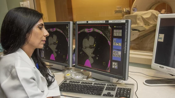
While cardiac ultrasound is the widely used imaging modality for heart assessments, computed tomography (CT), magnetic resonance imaging (MRI) and nuclear imaging are also used and are often complimentary, each offering specific details about the heart other modalities cannot. For this reason the clinical question being asked often determines the imaging test that will be used.

Thumbnail

Seeing the Future–in 3D: New CPT Codes Set the Stage for Expanding 3D Printing in Cardiology

Until recently, cardiologists’ eyes tended to glaze over at the mention of using 3D printing in their practices. Most believed the costs would be too high for routine use, that the applications and the price tag were better suited to academic applications.

Thumbnail

How should physicians treat STEMI patients with COVID-19? A new study aims to find out

A new international research study will focus on treatment strategies and outcomes of ST-segment elevation myocardial infarction (STEMI) patients with known or suspected COVID-19.

Thumbnail

‘A win-win for everyone’: Ultrasound technique provides improved arrhythmia assessments

A noninvasive ultrasound imaging technique can help healthcare providers localize atrial and ventricular cardiac arrhythmias, according to new research published in Science Translational Medicine. Could this method be added to clinical workflows in the near future?

What the COVID-19 pandemic means for cath labs

The COVID-19 pandemic has changed the practice of cardiology as we know it, leading to supplies shortages, delayed procedures, and an at-risk patient population fearful of the road ahead. How has the outbreak affected cath labs?

Thumbnail

ASNC monitoring potential Mo-99 supply shortages due to COVID-19

The president of the American Society of Nuclear Cardiology said there are no shortfalls reported at this time, but the organization is keeping an eye on the situation as more countries implement travel bans.

Thumbnail

What PET imaging can tell cardiologists about post-heart transplant survival

New research published in the Journal of Nuclear Medicine suggests PET myocardial perfusion imaging can be used to quantify myocardial blood flow and myocardial flow reserve, which have been proven as accurate indicators of adverse outcomes after heart transplants.

Thumbnail

‘Potentially everything we’ve hoped for’: Pulsed field ablation used for 1st time in U.S.

Pulsed field ablation technology has been used to treat atrial fibrillation for the first time in the U.S.

Thumbnail

Orsiro stent continues to outperform Xience at 3 years

Three-year data from the BIOFLOW-V trial, presented Feb. 23 at the 2020 CRT Congress in National Harbor, Md., reinforce the status of Biotronik’s Orsiro drug-eluting stent as superior to the popular Xience stent.

Around the web

Several key trends were evident at the Radiological Society of North America 2024 meeting, including new CT and MR technology and evolving adoption of artificial intelligence.

Ron Blankstein, MD, professor of radiology, Harvard Medical School, explains the use of artificial intelligence to detect heart disease in non-cardiac CT exams.