Cardiac Imaging

While cardiac ultrasound is the widely used imaging modality for heart assessments, computed tomography (CT), magnetic resonance imaging (MRI) and nuclear imaging are also used and are often complimentary, each offering specific details about the heart other modalities cannot. For this reason the clinical question being asked often determines the imaging test that will be used.

CT combo may be as effective as invasive testing for CAD

The combination of CT angiography (CTA) and CT myocardial perfusion (CTP) accurately identified patients with flow-limiting coronary artery disease (CAD) defined by 50 percent stenosis or greater using invasive tests, according to a study published online Nov. 19 in European Heart Journal.

Esaote showcases new prevention suite at EuroEcho 2013

This year’s EuroEcho-Imaging conference in Istanbul, Turkey (11-14 December) will see global medical imaging manufacturer, Esaote, showcase Prevention Suite: a unique package of ultrasound imaging modalities for both cardiac and vascular investigations. Prevention Suite provides clinicians with a wide range of tools for assessing a patient’s risk of cardiovascular disease quickly and reliably, often before symptoms occur. It is regarded as providing a ‘step change’ in terms of early detection and prevention of cardiovascular disease using ultrasound.

Thumbnail

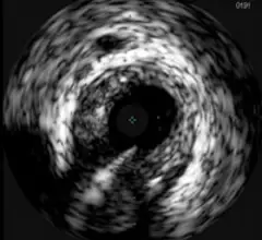
Panel outlines optimal use for IVUS, OCT & FFR

Clinicians should not rely on measurements obtained by intravascular ultrasound (IVUS) to evaluate the seriousness of non-left main coronary artery lesions without functional evidence to back them up, experts recommended in a consensus document published online Nov. 13 in Catheterization and Cardiovascular Interventions. This recommendation was one of several made for the optimal use of IVUS, optical coherence tomography (OCT) and fractional flow reserve (FFR).

CWRU team building an MRI-guided robotic heart catheter

In a matter of years, a doctor may see real-time images of a patient's beating heart and steer a robotic catheter through its chambers using the push and pull of magnetic fields while the patient lies inside a magnetic resonance imager.

World's largest imaging congress reveals latest techniques in heart failure

The latest science on the role of imaging in diagnosing and treating heart failure will be presented at EuroEcho-Imaging 2013, the official annual meeting of the European Association of Cardiovascular Imaging (EACVI), a registered branch of the European Society of Cardiology (ESC). It takes place during 11-14 December in Istanbul, Turkey, at the Istanbul Lutfi Kirdar Convention & Exhibition Centre (ICEC).

Left ventriculography use varies across VA hospitals

Veterans Affairs (VA) facilities vary in their use of left ventriculography and that variation cannot be explained by patient characteristics, according to a study published online Nov. 5 in Circulation: Cardiovascular Quality and Outcomes.

Thumbnail

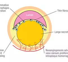
Radioisotope ID’s high-risk, ruptured coronary plaques

PET-CT using the radioisotope 18F-sodium fluoride (18F-NaF) identified high-risk and ruptured plaques in patients with stable and unstable coronary heart disease, according to a study published online Nov. 11 in The Lancet.

Thumbnail

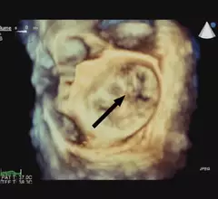
Vivid Valves: The Role of 3D Echo

Can 3D echocardiography transform any surgeon into a mitral valve repair expert?

Around the web

Several key trends were evident at the Radiological Society of North America 2024 meeting, including new CT and MR technology and evolving adoption of artificial intelligence.

Ron Blankstein, MD, professor of radiology, Harvard Medical School, explains the use of artificial intelligence to detect heart disease in non-cardiac CT exams.