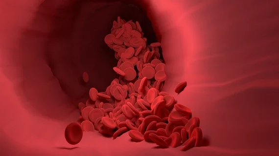
'Foaming' cells may help researchers evaluate CVD risk
Researchers have identified a specific gene expression in cells that may help predict a person's risk of developing cardiovascular disease (CVD), sharing their findings in Circulation.
“We started looking at macrophages, cells that eat lipids stuck to the walls of blood vessels,” corresponding author Beiyan Zhou, PhD, an immunologist at the UConn Health School of Medicine, said in a prepared statement.
These cells eat fatty deposits found on the artery walls—a process that Zhou said leaves them looking foamy, "like a clump of bubbles."
Macrophages are found in plaques along inflamed sections of blood vessels. This foamy plaque, the authors noted, is known to be associated with advanced CVD. Zhou et al. discovered that foamy macrophages consume fat that is in the wrong place. If macrophages are prevented from eating the fat, there is a higher risk of CVD. Patients with diabetes and autoimmune disease are at an even higher risk, according to the authors.
“Foaming is not necessarily bad; this is the first time that we can distinguish good and bad foaming," Zhou said. "It is the bad foaming that can cause heart attack."
The team hopes its research can help cardiologists identify patients facing an increased risk of CVD.
Through a collaboration with cardiologists at UConn Health, they have even developed a new risk prediction model that can determine a patient's risk of a heart attack or stroke within the next five years. That prediction model, they noted, outperformed many of the current methods specialists use to evaluate CVD risk.
Read the full study here.
