
PFA linked to increased risk of phrenic nerve damage
Phrenic nerve injury (PNI) may still be a significant concern following pulsed field ablation (PFA) for atrial fibrillation (AFib), according to a new single-center study published in Heart Rhythm.[1]
“PNI is a known complication of catheter ablation for AFib using thermal energies,” wrote first author Louis Chehirlian, MD, a cardiologist with CHU Timone in France, and colleagues. “Recently, PFA has emerged and given the theoretical tissue specificity of the lesions, it was expected that PNI would disappear. However, several publications have reported PNI cases, including persistent cases at hospital discharge.”
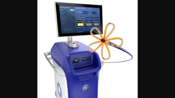
Chehirlian et al. tracked data from 64 adult AFib patients who underwent PFA with Boston Scientific’s Farapulse PFA system at a single facility. The mean patient age was 66.1 years old, 78.1% were men and the mean time since AFib diagnosis was 37.2 months. A majority (60.9%) of patients presented with persistent AFib, and the remaining patients presented with paroxysmal AFib.
Each patient underwent a transesophageal and transthoracic echocardiograph prior to treatment. Sequential compound motor action potential (CMAP) monitoring was used to monitor each patient for signs of PNI and measured using peak-to-peak millivolts. Right phrenic nerve monitoring, but not left phrenic nerve monitoring, was performed on each patient.
The mean operating time was 63.4 minutes, mean fluoroscopy time was 15 minutes and the most common catheter size (60.9%) was 31 mm.
“Three distinct patterns of diaphragmatic CMAP evolution were observed during the procedure,” the authors wrote. “The first pattern consisted of a stable CMAP amplitude, indicating the absence of PNI. In other cases, PNI occurred, resulting in a transient phase of complete signal loss lasting from a few seconds to several minutes (mean duration: 344.0 ± 298.0 seconds). This was consistently followed by a rapid recovery phase (1-2 minutes), during which CMAP amplitude returned either to its baseline value (second pattern) or to a lower value (third pattern). Following this rapid recovery phase, CMAP amplitude reached a plateau.”
The group added that diaphragmatic paralysis occurred in 40.6% of patients. The event rates were 80.8% during applications targeting the right superior pulmonary vein, 11.6% during applications targeting the right inferior pulmonary vein and 7.7% during applications targeting both veins and 7.7% during applications targeting the superior vena cava.
In addition, 65.4% of patients experienced an “abrupt onset of paralysis,” but the decrease in CMAP amplitude was more gradual for all other patients.
Four other key takeaways from the team’s research:
- Complete CMAP recovery was observed in 21.9% of patients.
- The duration of complete diaphragmatic paralysis was significantly longer in patients without a full recovery. This was the one patient factor linked to such a relationship.
- Patients with a lower body weight were more likely to face a risk of diaphragmatic paralysis after PFA.
- Incomplete diaphragmatic paralysis at hospital discharge was 24%, “which represents a notably high rate.”
“PNI appears to be a frequent event during PFA for AFib and often persists in an incomplete form at the end of the procedure, as evidenced by CMAP amplitude reduction,” the authors wrote. “Post-procedural fluoroscopy frequently reveals persistent diaphragmatic dysfunction. In one patient, fluoroscopic abnormality were still present at 3-month follow-up. These observations challenge the notion that PFA eliminates the risk of phrenic nerve damage and highlight the limitations of current standard assessment tools.”
They also emphasized that post-PFA fluoroscopy played a crucial role in identifying patients with PNI. Chest X-ray may be used instead in some facilities, but it may be “insufficient.”
“We believe that any iatrogenic complication should be reported,” the authors wrote. “No patient expects to be discharged from hospital with a new diaphragmatic deficit without being informed. It is the physician’s responsibility to identify such complications and to make every effort to prevent them.”
This study did have multiple limitations, including its small sample size and the fact that left phrenic nerve monitoring was not performed. In addition, the group noted, some patients treated early on during this trial did not receive follow-up fluoroscopy, so the prevalence of left-sided dysfunction “may be underestimated.”
To learn more, the authors recommend large-scale prospective studies specifically designed to track PNI after PFA.
Click here to read the full analysis.

Michael has more than 19 years of experience as a professional writer and editor. He has written at length about cardiology, radiology, artificial intelligence and other key healthcare topics.