Pre-procedure CT imaging benefits LAA occlusion in Henry Ford study
There is not agreement among some of the top structural heart experts who perform left atrial appendage occlusion (LAAO) if cardiac computed tomography (CT) is necessary to plan the procedure. Some experts at the 2022 Transcatheter Valve Therapies (TVT) Structural Heart Summit said in LAA sessions they used transesophageal echo (TEE), others use intracardiac echo (ICE) and others prefer CT. While there were several benefits to CT pointed out in sessions, reservations remained with some because of the cost and lack of reimbursement.
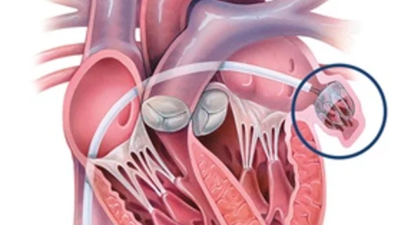
Recent findings from a Henry Ford Hospital Center for Structural Heart Disease study published in the Journal of American Heart Association showed CT use was associated with higher successful device implantation rates, shorter procedural times, and less frequent changes in device sizes.[1] This study looked at use of the self-expanding Watchman device during a catheter-based, minimally invasive procedure. Once implanted, the device permanently seals off that heart muscle pocket, which is the source of most stroke-causing clots in AFib patients.
Transcatheter LAA occlusion can reduce the risk of blood clots that can lead to stroke and eliminate the need to take anticoagulants for patients with non-valvular atrial fibrillation (AFib). However, the LAA comes in a variety of shapes, sizes and depths so it is important to image the LAA and properly size the device needed and to ensure it will fit in the LAA and not embolize.
“The standard method for imaging the heart to guide LAAO procedures is 2D TEE. This study aimed to assess the value of adding three dimensional CT imaging to that process, versus using only TEE imaging," explained Dee Dee Wang, MD, director of structural heart imaging at Henry Ford Hospital and the study’s senior author, in a statement. "Our findings indicate significant benefit by adding CT imaging, which uses X-ray to help create a more comprehensive 3D image of the heart.”
The hospital has been a pioneer in using advanced CT for structural heart procedure imaging, including transcatheter aortic valve replacement (TAVR), mitral valve repair and replacement (TMVR) and tricuspid valve repair and replacement (TTVR).
“CT imaging allows us to take all guesswork out of device implantation. We know that we can safely close the appendage and have a success of 98% when imaging is available,” said William O’Neill, MD, director of the Henry Ford Center for Structural Disease in the statement.
He said the 3D model helps structural heart interventional cardiologists choose a device that fits the patient’s unique LAA anatomy. It also helps with pre-planning to know where to make a transeptal puncture so the catheter will have the proper angle to access the LAA in the left atrium, making the procedure safer and and faster.
Study researchers retrospectively reviewed all LAAO procedures using the Watchman performed at a single center from May 2015 to December 2019. During this time, a total of 485 LAAOs were performed, including 328 (67.6%) cases who underwent additional CT for preprocedural planning and 157 (32.4%) cases using stand‐alone TEE for guidance.
Patients who had additive CT for preprocedural planning had a significantly higher rate of successful device implantation with <5 mm peri‐device leak than using stand‐alone TEE (98.5% versus 94.9%). Total procedural time was shorter in the additive CT group than the stand‐alone TEE group (median, 45.5 minutes versus 51.0 minutes). It was also less common to change device size after initial deployment in the additive CT group than the stand‐alone TEE group (5.6% versus 12.1%) with significantly more device upsizing in the stand‐alone TEE group (4% versus 9.4%).
Henry Ford now uses the newer FDA cleared Watchman FLX and the Amplatzer Amulet Left Atrial Appendage Occluder.
Related LAAO Content:
VIDEO: Advances in left atrial appendage occlusion technology — Interview with Devi Nair, MD
Amulet vs. Watchman: LAA occluder devices compared in new head-to-head trial
Abbott’s LAA closure solution for AFib patients gains FDA approval
What peridevice leaks after LAAO mean for patient health
Key interventional cardiology takeaways from ACC.22
LAAO outcomes significantly worse among women
Confirmed: Watchman FLX LAAC device safe for nonvalvular AFib patients
Cardiologist makes history, completes LAAO with new 3D ICE catheter
Reference:

