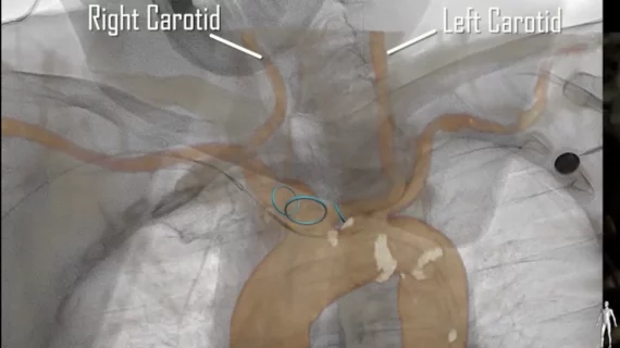
Cardiologists and surgeons perform first TAVR using CT imaging guidance on HF patient with blood clot
A team of cardiologists and surgeons at Hackensack University Medical Center in New Jersey believe they have made history by performing the world’s first transcatheter aortic valve replacement (TAVR) procedure guided by computed tomography (CT) fusion imaging on a patient with heart failure and a blood clot. The group shared its findings in JACC: Cardiovascular Interventions.[1]
The 78-year-old patient had a history of obesity, high blood pressure and coronary artery disease (CAD). He had previously been hospitalized for fainting. His left ventricular ejection fraction was 45% to 50%, and cardiac catheterization revealed that his CAD would be an issue for any minimally invasive procedures.
The care team decided that the best treatment option was TAVR with CT fusion imaging (CTFI) guidance and cerebral embolic protection.
“CTFI requires high-quality CT acquisition and creating a 3D heart model using a CT post-processing technique called segmentation on special software,” co-author Vladimir Jelnin, MD, Hackensack’s director of structural heart CT imaging, said in a statement. “This 3D model is then married to fluoroscopy using a process called coregistration, which projects the model on fluoroscopy at appropriate scale and orientation.”
“Fusion imaging is extremely helpful in identifying the cardiac structures during catheter and wire manipulation, especially in complex structural heart interventions,” added co-author Yuriy Dudiy, MD, a cardiac surgeon at Hackensack.
The team reported that the procedure was a success. He was discharged within a few days and had a positive one-month follow-up appointment.
“We believe this case is potentially groundbreaking because of its successful outcome and the fact that the presence of left-ventricle thrombus or blood clot has historically been considered to be a contraindication to TAVR, an alternative to open-heart surgery to replace heart valves in patients with heart disease,” lead author Rahul Vasudev, MD, a Hackensack interventional cardiologist, said in the same statement. “Although the nature of catheter manipulation in the left ventricle during the procedure cannot guarantee absence of contact with the blood clot, advances in imaging technology and embolic protection may allow transcatheter aortic valve replacement, if a surgical alternative is not possible, to be performed with greater safety in this setting.”
Related TAVR Content:
Transcaval TAVR outperforms transaxillary TAVR when femoral access is not an option
TAVR outcomes unaffected when women require a smaller valve prosthesis
The latest data on mitral valve infective endocarditis after TAVR
VIDEO: TAVR durability outperforms surgical valves
TAVR vs. surgery, FFR-guided PCI and DCB safety: Day 3 at ACC.22
Reference:

