Computed Tomography

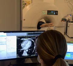
Computed tomography (CT) is a fast and accurate imaging modality often used in emergency settings and trauma imaging. CT scans, with or without (or both) iodinated contrast are frequently used to image the brain, chest, abdomen and pelvis, but also have post-imaging reconstructive capabilities for detailed orthopedic imaging. It is now a standard imaging modality in emergency rooms to quickly assess patients. CT uses a series of X-ray images shot as the gantry rotates around the patient. Computer technology assembles these into into a dataset volume than can be slices on any access, or advanced visualization software can extract specific parts of the anatomy for study. Find more content specific to cardiac CT.
CMS could increase CCTA payments—the American College of Cardiology wants to help

CMS wants feedback after proposing a significant update to CCTA reimbursement payments. The American College of Cardiology, a longtime champion for improved cardiac CT payments, is now helping its members make their voices heard.

Ron Blankstein, MD, FACC, FASNC, MSCCT, FASPC, associate director, cardiovascular imaging program, director, cardiac computed tomography, Brigham and Women’s Hospital, and a professor of medicine and radiology, Harvard Medical School, explains why Medicare is proposing increased coronary computed tomography angiography (CCTA) payments to hospitals.

Higher payments for CCTA could bring considerable change to cardiology

Ron Blankstein, MD, explains how and why Medicare is proposing an increase to hospital payments for coronary CT angiography.

CMS may double Medicare payments for cardiac CT

The agency is gathering feedback on a proposal to double the amount hospitals are paid for coronary computed tomography angiography, with the comment period ending Sept. 9.

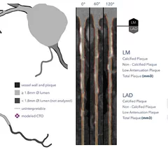
HeartFlow introduced its next generation artificial intelligence (AI) Plaque Analysis with an interactive experience at SCCT 2024. It shows a 3D plaque model and analysis by territory across calcified, non-calcified and low-attenuation plaques. This includes viewing cross-sectional, color-coded images of each plaque type where it was quantified along the vessel. #SCCT #SCCT24 #SCCT2024

Updated HeartFlow technology offers interactive features, full integration with FFR-CT 

HeartFlow is using SCCT 2024 to introduce the world to its updated Plaque Analysis platform. “Accurately diagnosing a patient’s risk for coronary artery disease is critical for determining the best treatment," Chief Medical Officer Campbell Rogers, MD, explained.

Peter Monteleone, MD, an interventional cardiologist, national director of cardiovascular research at Ascension Health, and assistant professor, UT Austin Dell School of Medicine, explained the use of artificial intelligence (AI) to independently identify an emergency stroke or pulmonary embolism (PE) finding on a CT scan and automatically alert critical care team members. His health system uses this type of AI for earlier activation of the pulmonary embolism response team (PERT).

AI critical care software revolutionizes emergency response

Ascension Health in Texas uses AI that can read CT scans for stroke and pulmonary embolism to activate care teams before the images even get into the PACS.

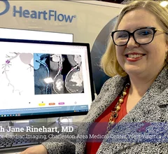
Sarah Jane Rinehart, MD, director of cardiac imaging, Charleston Area Medical Center, Charleston West Virginia, as been using the FDA-cleared RoadMap artificial intelligence algorithm from HeartFlow in studies and in clinical used since it was cleared and said it helps cardiologists in several ways. #ACC #ACC24 #ACC2024 #Heartflow #AIhealth

AI improves CT assessments, boosts care for real-world heart patients

Automated AI-generated measurements combined with annotated CT images can improve treatment planning and help referring physicians and patients better understand their disease, explained Sarah Jane Rinehart, MD, director of cardiac imaging with Charleston Area Medical Center.

Advanced artificial intelligence (AI) models can evaluate cardiovascular risk in routine chest CT scans without contrast, according to new research published in Nature Communications.[1] In fact, the authors noted, the AI approach may be more effective at identifying issues than relying on guidance from radiologists.

AI predicts cardiovascular risk during CT scans—no invasive tests or contrast required

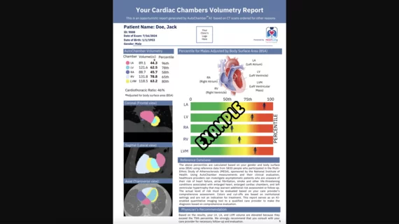
Two advanced algorithms—one for CAC scores and another for segmenting cardiac chamber volumes—outperformed radiologists when assessing low-dose chest CT scans. 

Medicare drops AUC requirement for advanced imaging, ASNC celebrates

"This was an unneeded burden, which was solely adding to the administrative hassles of medicine," said American Society of Nuclear Cardiology President Larry Phillips.

Around the web

Ron Blankstein, MD, professor of radiology, Harvard Medical School, explains the use of artificial intelligence to detect heart disease in non-cardiac CT exams.

Eleven medical societies have signed on to a consensus statement aimed at standardizing imaging for suspected cardiovascular infections.