Computed Tomography

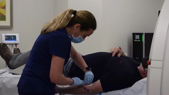
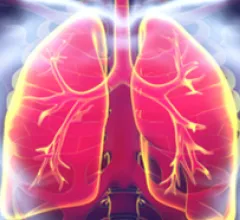
Computed tomography (CT) is a fast and accurate imaging modality often used in emergency settings and trauma imaging. CT scans, with or without (or both) iodinated contrast are frequently used to image the brain, chest, abdomen and pelvis, but also have post-imaging reconstructive capabilities for detailed orthopedic imaging. It is now a standard imaging modality in emergency rooms to quickly assess patients. CT uses a series of X-ray images shot as the gantry rotates around the patient. Computer technology assembles these into into a dataset volume than can be slices on any access, or advanced visualization software can extract specific parts of the anatomy for study. Find more content specific to cardiac CT.
Leslee Shaw, PhD, and former presidents of both SCCT and ASNC discusses the role of CT and FFR-CT in the 2021 chest pain guidelines.

VIDEO: Cardiac CT now recommended as a front-line chest pain assessment tool

Leslee Shaw, PhD, director of the Blavatnik Family Women’s Health Research Institute at Mount Sinai Hospital in New York and former president of the Society of Cardiovascular Computed Tomography (SCCT) and the American Society of Nuclear Cardiology (ASNC), explains the role of cardiac computed tomography (CT) in the recent 2021 chest pain guidelines.

The ASNC is one of several medical imaging societies asking Congress to repeal the appropriate use criteria (AUC) criteria mandate. They say it poses issues for clinicians and is becoming outdated by changes in CMS payment systems. The AUC requirements call for documentation using CVMS authorized software in order to show advanced imaging such as nuclear and CT is justified, or else Medicare payments might be withheld.

American Society of Nuclear Cardiology urges Congress to speed prior authorizations, repeal AUC mandate 

Over the past few weeks, members of ASNC’s Health Policy Committee have held meetings with their members of Congress.

AI model able to ID early signs of type 2 diabetes on imaging results

The authors hope their findings could lead to earlier diagnoses and improvements in patient care. 

chest pain lung pulmonary embolism

Image quality is not an issue for AI model that detects pulmonary embolisms on CT

CTPA is the standard of care for diagnosing PE, but suboptimal scans make it difficult to reach a diagnosis. A new Clinical Imaging study tests the effectiveness of AI when image quality is lacking.

Thumbnail

Automated CT scoring system accurately predicts prognosis in stroke patients

The study used non-contrast CT and CT perfusion imaging to analyze agreement between an automated reader and human radiologists with differing experience levels.

Thumbnail

‘A watershed moment’: HeartFlow praises new chest pain guidelines

The company showed its support for the guidelines in a new statement. 

Thumbnail

Radiologists should watch for these 3 pulmonary findings linked to increased COVID mortality

Experts looked beyond common pulmonary consolidations, finding a handful of accurate indicators of in-hospital mortality.

Thumbnail

Negative CT-PA scans could increase time-to-disposition in patients seen for pulmonary embolism

Patient and environmental factors proved not to be major culprits but scan results were key, experts reported in BMC Emergency Medicine.

Around the web

Ron Blankstein, MD, professor of radiology, Harvard Medical School, explains the use of artificial intelligence to detect heart disease in non-cardiac CT exams.

Eleven medical societies have signed on to a consensus statement aimed at standardizing imaging for suspected cardiovascular infections.

Trimed Popup
Trimed Popup