Computed Tomography

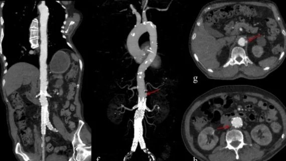
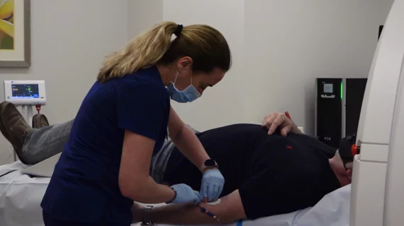
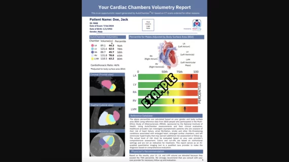
Computed tomography (CT) is a fast and accurate imaging modality often used in emergency settings and trauma imaging. CT scans, with or without (or both) iodinated contrast are frequently used to image the brain, chest, abdomen and pelvis, but also have post-imaging reconstructive capabilities for detailed orthopedic imaging. It is now a standard imaging modality in emergency rooms to quickly assess patients. CT uses a series of X-ray images shot as the gantry rotates around the patient. Computer technology assembles these into into a dataset volume than can be slices on any access, or advanced visualization software can extract specific parts of the anatomy for study. Find more content specific to cardiac CT.

Thumbnail

Document offers guidance on curbing radiation exposure during CV imaging

An expert consensus document published May 2 in the Journal of the American College of Cardiology offers recommendations for limiting radiation exposure to both patients and medical personnel during cardiovascular imaging procedures.

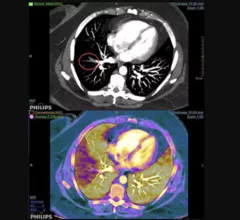
Example of spectral, or dual-energy CT, confirming a pulmonary embolism (PE). Image courtesy of Philips Healthcare

MRA vs CTA for evaluating pulmonary embolism: Does the chosen modality impact downstream imaging utilization?

MR angiography (MRA) is a relatively new alternative to CT angiography (CTA) for the evaluation of suspected pulmonary embolism (PE) and it even has one major advantage over CTA: it does not expose patients to ionizing radiation.

Thumbnail

James K. Min named editor-in-chief of Journal of Cardiovascular Computed Tomography

James K. Min, MD, has been chosen to be the new editor-in-chief of the Journal of Cardiovascular Computed Tomography (JCCT), the official journal of the Society of Cardiovascular Computed Tomography (SCCT). He will serve a five-year term.

James Min named editor-in-chief of Journal of Cardiovascular Computed Tomography

Vienna, VA (April 25, 2017) — James K. Min, MD, FSCCT, has been selected for a five-year term as the new editor-in-chief of the Journal of Cardiovascular Computed Tomography (JCCT). He in turn has named Todd C. Villines, MD, FSCCT, as executive editor along with Gudrun Feuchtner, MD.

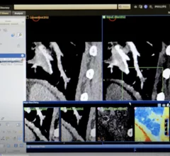
Example of pulmonary embolism (PE) detection on CT and confirmed using spectral CT. Image courtesy of Philips Healthcare

Researchers 'concerned' CT angiography topped MRA in follow-ups for pulmonary embolism

A recent study found whether a patient received MR angiography (MRA) instead of CT angiography (CTA) for a pulmonary embolism (PE) had little effect on the rate of follow-up chest CT or MRI one year later.

Thumbnail

Non-hospital-based facilities offering cardiac imaging services a rarity in Massachusetts

In 2017, Anthem implemented a new policy that requires outpatient MR and CT scans not considered medically necessary to be completed at a freestanding imaging facility to receive coverage. This went into effect in several states, sparking its fair share of controversy along the way, and a team of Boston-based researchers wanted to know how such a policy could impact patient care in Massachusetts.

Thumbnail

Chest CT for suspected pulmonary embolism in the ED: Utilization is up, diagnostic yield is down

CT utilization in the emergency department (ED) has increased significantly in the last few decades, a fact often cited as one of the major reasons healthcare costs and radiation exposure are on the rise. So what can be done to reverse that trend?

Noted cardiologist Jagat Narula receives SCCT award

Jagat Narula, MD, PhD, received the Arthur S. Agatston Cardiovascular Disease Prevention award from the Society of Cardiovascular Computed Tomography (SCCT).

Around the web

Ron Blankstein, MD, professor of radiology, Harvard Medical School, explains the use of artificial intelligence to detect heart disease in non-cardiac CT exams.

Eleven medical societies have signed on to a consensus statement aimed at standardizing imaging for suspected cardiovascular infections.

Trimed Popup
Trimed Popup