Computed Tomography

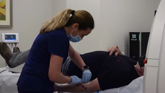
Computed tomography (CT) is a fast and accurate imaging modality often used in emergency settings and trauma imaging. CT scans, with or without (or both) iodinated contrast are frequently used to image the brain, chest, abdomen and pelvis, but also have post-imaging reconstructive capabilities for detailed orthopedic imaging. It is now a standard imaging modality in emergency rooms to quickly assess patients. CT uses a series of X-ray images shot as the gantry rotates around the patient. Computer technology assembles these into into a dataset volume than can be slices on any access, or advanced visualization software can extract specific parts of the anatomy for study. Find more content specific to cardiac CT.
Fasting before contrast-enhanced CT exams is unnecessary, experts say

In some instances, not eating solid foods can actually make patients feel sick, leading to nausea and dehydration, researchers explained recently.

Thumbnail

Offering imaging exam for free ups utilization by 546%, with key gains in underserved populations

University Hospitals Health System tested the pricing change in a bid to boost outcomes in its service area. 

Thumbnail

Expert panel recommends coronary CTA as first choice when evaluating for stable CAD

The American College of Cardiology Summit on Technology Advances in Coronary Computed Tomography Angiography shared its assessment in a new report. 

Aidoc announces CE mark for the first AI-based workflow tool for Pulmonary Embolism

Aidoc’s solution for flagging and prioritizing pulmonary embolisms is now commercially available in Europe

Thumbnail

Plaque characteristics boost predictive power of CTA risk scoring

A CT angiography (CTA)-derived score that also incorporated the extent, location and composition of coronary plaque outperformed a model that focused only on the severity of stenosis, researchers reported Jan. 16 in JACC: Cardiovascular Imaging.

Thumbnail

Industry group releases new guidelines for CT use in TAVR procedures

The Society of Cardiovascular Computed Tomography (SCCT) released updated guidelines for using CT imaging in patients undergoing transcatheter aortic valve replacement (TAVR).

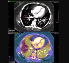
Example of spectral, or dual-energy CT, confirming a pulmonary embolism (PE). Image courtesy of Philips Healthcare

Dual-energy CTA iodine maps offer small benefit in diagnosing pulmonary embolism

Dual-energy (DE) CT iodine maps offer a slight benefit when paired with traditional CT angiography images in diagnosing pulmonary embolism (PE), reported researchers in a Sept. 11 Radiology study.

Han awarded the inaugural 2018 DeHaan Award for Innovation in Cardiology

Vienna, VA (August 13, 2018) — B. Kelly Han, MD, is the recipient of the inaugural 2018 DeHaan Award for Innovation in Cardiology, which includes a $200,000 grant.

Around the web

Ron Blankstein, MD, professor of radiology, Harvard Medical School, explains the use of artificial intelligence to detect heart disease in non-cardiac CT exams.

Eleven medical societies have signed on to a consensus statement aimed at standardizing imaging for suspected cardiovascular infections.

Trimed Popup
Trimed Popup