Computed Tomography

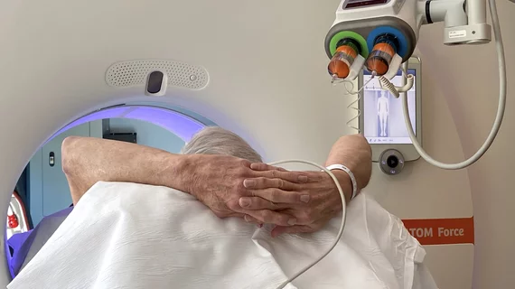
Computed tomography (CT) is a fast and accurate imaging modality often used in emergency settings and trauma imaging. CT scans, with or without (or both) iodinated contrast are frequently used to image the brain, chest, abdomen and pelvis, but also have post-imaging reconstructive capabilities for detailed orthopedic imaging. It is now a standard imaging modality in emergency rooms to quickly assess patients. CT uses a series of X-ray images shot as the gantry rotates around the patient. Computer technology assembles these into into a dataset volume than can be slices on any access, or advanced visualization software can extract specific parts of the anatomy for study. Find more content specific to cardiac CT.


VIDEO: Cardiac CT as a front-line chest pain exam in the ED

Harold Litt, MD, explains the use of cardiac CT as a primary imaging exam for chest pain at his emergency department over the past decade.

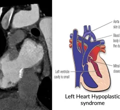
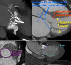
CT imaging showing the congenital heart defect of left heart hypoplastic syndrome.

VIDEO: Congenital heart imaging with cardiac CT

As the population of patients with adult congenital heart disease grows, they are presenting to adult cardiology clinics and being imaged with CT. Many also do not have access or cannot be imaged by MRI, said Renee Bullock-Palmer, MD.

New recommendations for cardiac CT in cardio-oncology imaging

The statement was written to fill gaps in recommendations from prior consensus statements and guidelines in regards to the use of CCT in cardio-oncology, including use of calcium scoring and ruling out coronary disease when cardiac function is impaired.

Examples of structural heart transcatheter valve replacement procedure planning CT scans and post procedure followup for TMVR and TAVR.

VIDEO: CT imaging for TAVR and TMVR structural heart interventions

Joao Cavalcante, MD, director, cardiac MRI and structural CT labs, Minneapolis Heart Institute, discusses the use of cardiac CT imaging to plan and guide structural heart procedures. 

VIDEO: Gender differences in women with cardiovascular disease and implications for imagers

Erin D. Michos, MD, co-editor in chief of the American Journal of Preventive Cardiology, discusses gender differences in heart disease presentations.

Automated CT body composition analysis predicts risk of stroke and heart attack

Researchers observed visceral fat area (VFA) measurements derived from abdominal CT scans to be associated with increased cardiovascular risk. 

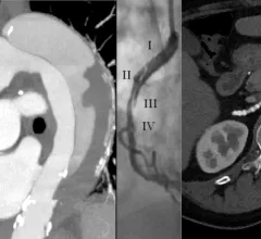
Vascular CT beyond the coronaries includes imaging for aortic aneurism, aortic dissection, SCAD and fibromuscular dysplasia. #SCCT #SCCT2022 #SCAD #yescct

VIDEO: Vascular imaging applications for CT beyond the heart

Society of Cardiovascular Computed Tomography (SCCT) President Brian Ghoshhajra, MD, explains the vascular imaging sessions at SCCT 2022 that went beyond the usual coronaries and structural heart evaluations.

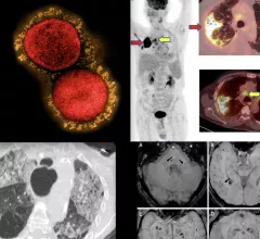
COVID-19 medical imaging examples of various clinical presentations. SARS-CoV-2 clinical imaging presentations.

PHOTO GALLERY: What does COVID-19 look like on medical imaging?

This image gallery shows what the various clinical presentations associated with the COVID-19 virus that have been documented during the coronavirus pandemic.

Around the web

GE HealthCare said the price of iodine contrast increased by more than 200% between 2017 to 2023. Will new Chinese tariffs drive costs even higher?

Several key trends were evident at the Radiological Society of North America 2024 meeting, including new CT and MR technology and evolving adoption of artificial intelligence.