Computed Tomography

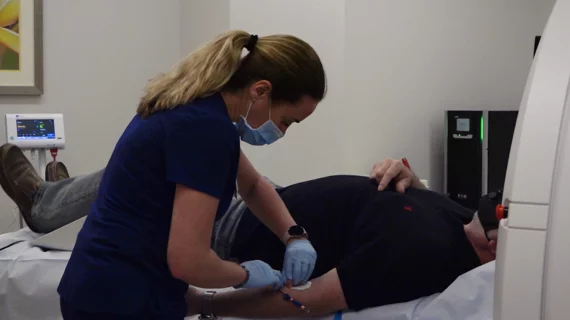
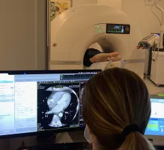
Computed tomography (CT) is a fast and accurate imaging modality often used in emergency settings and trauma imaging. CT scans, with or without (or both) iodinated contrast are frequently used to image the brain, chest, abdomen and pelvis, but also have post-imaging reconstructive capabilities for detailed orthopedic imaging. It is now a standard imaging modality in emergency rooms to quickly assess patients. CT uses a series of X-ray images shot as the gantry rotates around the patient. Computer technology assembles these into into a dataset volume than can be slices on any access, or advanced visualization software can extract specific parts of the anatomy for study. Find more content specific to cardiac CT.
Thumbnail

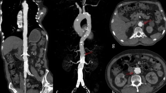
Coronary artery calcification often unreported in CT angiography studies

Coronary artery calcification (CAC) is often not reported in pulmonary CT angiography studies, requiring CAC assessment by radiologists in demographics at risk, according to a study published in the April issue of the American Journal of Roentgenology. 

Radiology: Decision support reins in CT pulmonary angiographic exams

Evidence-based decision support programs may be able to reduce the number of unnecessary CT pulmonary angiographic exams used to evaluate pulmonary emboli in the emergency department, according to a study published online Dec. 20 in Radiology.

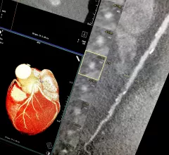
Example of a curved MPR image reconstruction of entire length of of a coronary artery on a cardiac CT scan to better show calcified and soft plaque burden inside the vessel. The thumbnail dots on the left side of the image are cross sectional views of the vessel. Siemens example on the expo floor.

The CCTA Playbook: A Guide to Coding, Reimbursement and Operations

A compendium of the business intelligence required to launch a CCTA service

Be Mindful of the Stark Law in CCTA Alliances

Numerous radiology groups are currently discussing arrangements whereby cardiologists and the radiologists would essentially split the responsibilities for reading coronary CT angiography (CCTA) studies. Most commonly, as proposed, the cardiologists would read and generate a signed interpretation report, but limited to the cardiac portion of the

Around the web

Ron Blankstein, MD, professor of radiology, Harvard Medical School, explains the use of artificial intelligence to detect heart disease in non-cardiac CT exams.

Eleven medical societies have signed on to a consensus statement aimed at standardizing imaging for suspected cardiovascular infections.

Trimed Popup
Trimed Popup