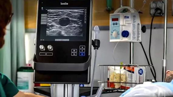
Ultrasound guidance for femoral access did not reduce bleeding or vascular complications in TCT late-breaker
In one of the largest multicenter randomized trials comparing ultrasound with fluoroscopic guidance vs. fluoroscopic guidance alone to guide femoral artery vascular access for patients undergoing cardiac procedures, many were surprised to find ultrasound did not reduce vascular complications and bleeding.
This was the result of the UNIVERSAL Trial presented at Transcatheter Cardiovascular Therapeutics (TCT) 2022 meeting, and simultaneously published in JAMA Cardiology.[1]
While the study found that ultrasound did not did not reduce vascular complications or bleeding, it did lower the number of attempts and venipuncture.
“Although ultrasonography guidance for femoral access did not reduce the primary events of bleeding or vascular complications, some benefits were identified,” said Sanjit S. Jolly, MD, MSc, interventional cardiologist with Hamilton Health Sciences and professor of medicine at McMaster University, said in a statement from the Cardiovascular Research Foundation (CRF). “Ultrasonography did improve first-pass success and reduced the number of attempts as well as the risk of venipuncture. Therefore, larger trials may be able to identify additional benefits for this technique.”
Prior to wide use of point-of-care ultrasound systems in the cath lab to guide needles into the artery to gain access, anatomical landmarks and angiography were used. It has widely been believed ultrasound would help guide vascular access much more precisely and prevent costly complications and extended length of stays. Jolly explained ultrasound still offers a lot of benefits, but it is possible more training is needed to improve how it is used when femoral access is needed.
“We need to put this into perspective because ultrasonography has no risks and is low cost, and so we need to focus on training. Finally, transradial access remains the best way to prevent femoral access bleeding," Jolly said.
Although transradial access for cardiac procedures reduces bleeding at the access site by more than 60%, femoral access is still needed for large-bore procedures and patients with small or occluded radial arteries. The rate of femoral vascular complications remains high in this subgroup of patients and previous randomized trials of ultrasound (US) guidance have shown mixed results.
Overall meta-analysis of ultrasound guided femoral access is still positive
It was noted in this study that its data was shared with the meta-analysis of all the available trials that examined ultrasound guidance. Including the UNIVERSAL study data, the PROSPERO international prospective registery of systematic reviews with more than 4,000 patients, shows that ultrasound does reduce major bleeding and vascular complications (RR 0.58; 95% CI 0.43-0.76).
Details on the UNIVERSAL trial on ultrasound vs. angiography for femoral access
Between June 26, 2018, and April 26, 2022, 621 patients were randomized 1:1 at two centers in Canada. Patients with ST-elevation myocardial infarction (STEMI) were not eligible to participate. The trial population had a high rate of comorbidities, including 51% of which had a prior myocardial infarction, 42% had diabetes, 45% had prior percutaneous coronary intervention (PCI), 57% had previous coronary bypass surgery, 19% had atrial fibrillation and 18% had peripheral vascular disease. For the index procedure, 80% were 6 French, 42% underwent PCI, 14% underwent chronic total occlusion (CTO) PCI and closure devices were used in 52.1% of patients.
The primary outcome, defined as the composite of major vascular complications or major bleeding based on Bleeding Academic Research Consortium (BARC) 2, 3 or 5 criteria within 30 days, occurred in 40 of 311 patients (12.9%) in the ultrasonography group versus 50 of 310 patients (16.1%) without ultrasonography (odds ratio, 0.77 [95% CI, 0.49-1.20]; p=.25). The key secondary outcome of the rates of BARC 2, 3, or 5 bleeding were 10.0% (31 of 311) versus 10.7% (33 of 310) (odds ratio, 0.93 [95% CI, 0.55-1.56]; p=.78). The rates of major vascular complications were 6.4% (20 of 311) vs 9.4% (29 of 310) (odds ratio, 0.67 [95% CI, 0.37-1.20]; p=.18).
The study also found that ultrasonography improved first-pass success (277 of 311 [86.6%] vs 222 of 310 [70.0%]; odds ratio, 2.76 [95% CI, 1.85-4.12]; p<.001) and reduced the number of arterial puncture attempts (mean [SD], 1.2 [0.5] vs 1.4 [0.8]; mean difference, −0.26 [95% CI, −0.37 to −0.16]; p< .001) and venipuncture (10 of 311 [3.1%] vs 37 of 310 [11.7%]; odds ratio, 0.24 [95% CI, 0.12-0.50]; p< .001) with similar times to access.
In the pre-specified subgroup analysis, patients who had a closure device had a significant benefit for ultrasound (OR 0.44 95% CI 0.23-0.82, interaction p=0.004). Jolly said this makes sense, since ultrasound avoids multiple punctures and allows the operator to choose a site free of disease or calcium.
The study was funded by Hamilton Health Sciences Foundation and McMaster Cardiology program.

