Cardiac Imaging

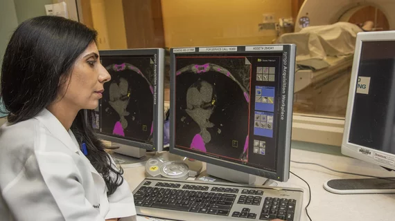
While cardiac ultrasound is the widely used imaging modality for heart assessments, computed tomography (CT), magnetic resonance imaging (MRI) and nuclear imaging are also used and are often complimentary, each offering specific details about the heart other modalities cannot. For this reason the clinical question being asked often determines the imaging test that will be used.

Thumbnail

Industry groups oppose proposed myocardial PET cuts

When CMS released the 2020 Medicare Physician Fee Schedule (MPFS) proposed rule, it included revisions that could lead to technical component payment reductions of up to 80% for certain services related to myocardial PET scans.

Thumbnail

Applying AI to ECG data helps gauge patients’ ‘physiologic age’

A study funded by the Mayo Clinic and published in Circulation: Arrhythmia and Electrophysiology this week suggests AI could be successfully applied to ECG data to measure a person’s overall health status.

Thumbnail

Echocardiography the most used imaging modality in HF patients

A study published in JAMA Network Open August 11 suggests that, despite the growing use of imaging modalities like computed tomography and MRI, echocardiography remains the most popular method for imaging patients with heart failure.

Thumbnail

Novel imaging approach IDs CAD without the risk of a contrast agent

Researchers at Beth Israel Deaconess Medical Center in Boston have developed an imaging technique that allows physicians to diagnose CAD without the risk of a contrast agent.

The economic impact of MR on heart failure patients

Newly diagnosed HF patients with concomitant mitral regurgitation can expect more admissions, longer hospital stays and pricier medical bills than HF patients without MR, according to an analysis published in the American Journal of Cardiology.

How researchers are using patient selfies to monitor blood pressure

A study published this week in Circulation: Cardiovascular Imaging details how one Canadian scientist and his team are using 2-minute video selfies to track patients’ blood pressure.

Thumbnail

Researchers 3D-print heart components using collagen

Researchers at Carnegie Mellon University’s College of Engineering have devised a way to 3D bioprint tissue scaffolds out of collagen, putting them one step closer to the goal of printing a full-sized, functional human heart.

Thumbnail

Medtronic, Viz.ai partner to improve LVO stroke detection

Medtronic and Viz.ai, an AI-driven tech company, have joined forces to promote the adoption of imaging software that could drastically cut the time it takes physicians to identify suspected large vessel occlusion.

Around the web

Several key trends were evident at the Radiological Society of North America 2024 meeting, including new CT and MR technology and evolving adoption of artificial intelligence.

Ron Blankstein, MD, professor of radiology, Harvard Medical School, explains the use of artificial intelligence to detect heart disease in non-cardiac CT exams.