Cardiac Imaging

While cardiac ultrasound is the widely used imaging modality for heart assessments, computed tomography (CT), magnetic resonance imaging (MRI) and nuclear imaging are also used and are often complimentary, each offering specific details about the heart other modalities cannot. For this reason the clinical question being asked often determines the imaging test that will be used.

Thumbnail

Intravascular ultrasound may provide better long-term outcomes in complex PCI

Intravascular ultrasound (IVUS)-guided PCI was associated with lower long-term risk of cardiac death than an angiography-guided approach in patients with complex lesions, according to a single-center study published in JACC: Cardiovascular Interventions.

Thumbnail

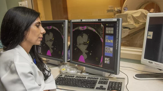
Radiologists often overlook cardiovascular warning signs during cancer imaging

Radiologists commonly overlook potentially important information about a patient’s heart health when they’re performing mammograms or CT scans before starting cancer treatment, according to research set to be presented at the American College of Cardiology’s scientific sessions March 16-18 in New Orleans.

Ultrasound societies want the FDA to remove contrast label warning

The International Contrast Ultrasound Society (ICUS) is again urging the FDA to remove the “black box” warning from ultrasound contrast agent (UCA) labels, following its filing of a September 2018 citizen petition with the federal agency.

Thumbnail

CT angiography trumps functional testing in diabetic patients with suspected CAD

A new analysis from the PROMISE trial suggests CT angiography (CTA) may be a superior front-line diagnostic test for patients with diabetes and symptoms suggestive of coronary artery disease (CAD).

Aidoc announces CE mark for the first AI-based workflow tool for Pulmonary Embolism

Aidoc’s solution for flagging and prioritizing pulmonary embolisms is now commercially available in Europe

Thumbnail

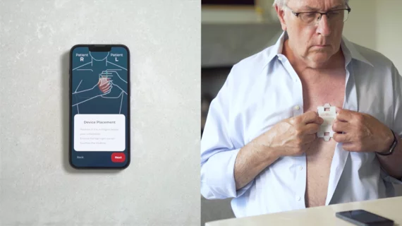
Chest strap ECG effectively detects AFib

Electrocardiograms (ECG) acquired using a chest strap could be a quality alternative to traditional ECGs when used to diagnose atrial fibrillation (AF), researchers reported in the American Journal of Cardiology.

Thumbnail

Cardiac MRI-derived T2 mapping may help heart failure patients

Researchers used T2 mapping taken from weekly cardiac MRIs to help identify cardiotoxicity at an early stage, according to results of a pig study published Feb. 18 in the Journal of the American College of Cardiology. The findings could help cancer patients at risk of chemo-induced heart failure.

Thumbnail

T2 mapping may uncover cardiotoxic marker early enough to prevent heart failure

T2 mapping derived from weekly cardiac MRIs helped researchers identify cardiotoxicity at an early and reversible stage, a finding which may have implications for cancer patients at risk of chemotherapy-induced heart failure.

Around the web

Several key trends were evident at the Radiological Society of North America 2024 meeting, including new CT and MR technology and evolving adoption of artificial intelligence.

Ron Blankstein, MD, professor of radiology, Harvard Medical School, explains the use of artificial intelligence to detect heart disease in non-cardiac CT exams.