Cardiac Imaging

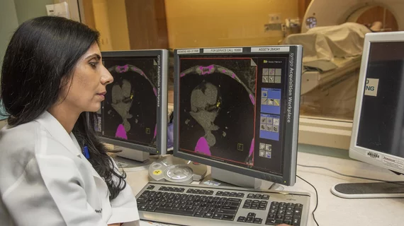
While cardiac ultrasound is the widely used imaging modality for heart assessments, computed tomography (CT), magnetic resonance imaging (MRI) and nuclear imaging are also used and are often complimentary, each offering specific details about the heart other modalities cannot. For this reason the clinical question being asked often determines the imaging test that will be used.

Thumbnail

CEO & HRS Leader’s Strategy: Listen, Ask, Recruit & Step Back

As an early career electrophysiologist, Richard I. Fogel, MD, learned to raise his hand when projects needed a captain. Now he’s CEO of the St. Vincent Medical Group in Indianapolis and president of the Heart Rhythm Society (HRS). His formula? Listen, ask good questions, surround yourself with talented people and let them do their jobs, he explains in a Q&A with Cardiovascular Business.

Thumbnail

FFR & IVUS: Putting Tools to Good Use

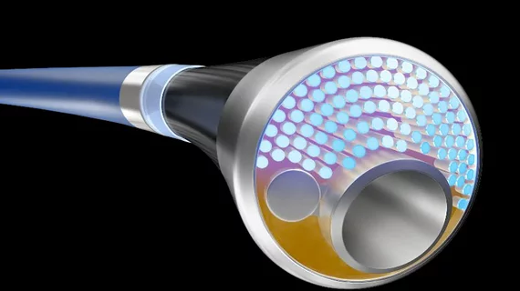
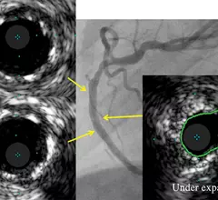
Think of fractional flow reserve (FFR) as a hex wrench. Or intravascular ultrasound (IVUS) as a screwdriver. Both imaging methods are highly useful in the right situation, according to physicians. Both methods can be highly effective, when used. However, there is some disagreement about where and when these tools should be applied.

Thumbnail

Stress Test: Evidence Mounts in Regadenoson’s Favor

The FDA issued a warning in late 2013 about rare but serious adverse events in some patients who received regadenoson, one of nuclear cardiology’s favorite pharmacologic stress agents. That didn’t dampen enthusiasm for it in the least. Instead, physicians show it has much more to offer. 

Thumbnail

Valves in 3D: Updated Echo Techniques Add Clarity

Three-dimensional imaging adds a needed perspective to cardiac procedures. Nowhere is this more pronounced than in the field of valvular interventions. 

Coronary CT angiography is cost-effective for patients with stable chest pain

For patients with stable chest pain, utilizing coronary CT angiography before cardiac stress imaging was cost-effective, according to a microsimulation model.

Thumbnail

A safer cath lab

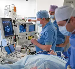
The cardiology community has heeded the call to protect patients from radiation exposure in the cath lab. Now it is time to focus on the operators and staff.

3D CMR shines as diagnostic tool for detecting CAD

Whole-heart 3D myocardial perfusion cardiac MR (CMR) accurately detected coronary artery disease (CAD) in a study that used fractional flow reserve as a reference, making CMR a possible contender to techniques that are invasive and expose patients to ionizing radiation.

VasCore Training & Education Center (VTEC) opens in downtown Boston

Today the founders of VasCore, The Vascular Ultrasound Core Laboratory, announce the opening of the VasCore Training & Education Center (VTEC).

Around the web

Several key trends were evident at the Radiological Society of North America 2024 meeting, including new CT and MR technology and evolving adoption of artificial intelligence.

Ron Blankstein, MD, professor of radiology, Harvard Medical School, explains the use of artificial intelligence to detect heart disease in non-cardiac CT exams.