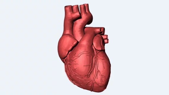
Personalized 3D model of hearts can help locate, treat arrhythmia
Scientists hope personalized data from patients experiencing irregular heartbeats will help improve accuracy in heart ablation procedures. The 3D simulations of 21 patients allow physicians to locate arrhythmia by “poking” the simulated heart with small electrical signals in various locations.
The researchers published their proof-of-concept study online Sept. 3 in Nature Biomedical Engineering.
"Cardiac ablation, or the destruction of tissue to stop errant electrical impulses, has been somewhat successful but hampered by a lot of guesswork and variability in the way that physicians figure out which locations to zap with a catheter," Natalia Trayanova, PhD, with the department of biomedical engineering at the Johns Hopkins University in Baltimore, said in a prepared statement. "Our new study results suggest we can remove a lot of the guesswork, standardize treatment and decrease the variability in outcomes, so that patients remain free of arrhythmia in the long term.”
The team used MRI to create personalized heart models of 21 patients who had undergone cardiac ablation for infarct-related ventricular tachycardia. They used the 3D simulation to guide ablation treatments for five patients—with two remained tachycardia free for 23 and 21 months, one remained symptom free after two months and two did not undergo ablation because the simulation showed tachycardias would not inducible.
The researchers hope predictions based on these MRI-created simulations could help predict outcomes, reduce complications and abbreviate the cardiac mapping process.

