Nuclear Medicine

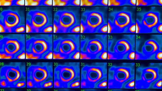
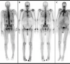
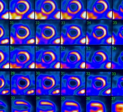
Nuclear medicine (also called molecular imaging) includes positron emission computed tomography (PET) and single photon emission computed tomography (SPECT) imaging. Nuclear imaging is achieved by injecting small amounts of radioactive material (radiopharmaceuticals) into patients before or during their scan. These can use sugars or chemical traits to bond to specific cells. The radioactive material is taken up by cells that consume the sugars. The radiation emitted from inside the body is detected by photon detectors outside the body. Computers take the data to assemble images of the radiation emissions. Nuclear images may appear fuzzy or ghostly rather than the sharper resolution from MRI and CT.  But, it provides metabolic information at a cellular level, showing if there are defects in the function of the heart, areas of very high metabolic activity associated with cancer cells, or areas of inflammation, data not available from other modalities. These noninvasive imaging exams are used to diagnose cancer, heart disease, Alzheimer’s and Parkinson’s disease, bone disorders and other disorders. 
Global shortage of nuclear imaging isotopes may be over

The nuclear imaging isotope shortage of molybdenum-99 may be over now that the sidelined reactor is restarting. ASNC's president says PET and new SPECT technologies helped cardiac imaging labs better weather the storm.

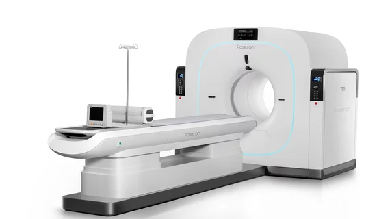
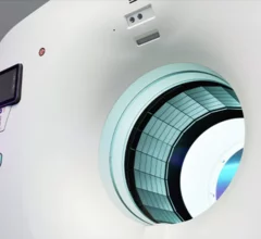
GE HealthCare Omni Legend PET/CT scanner production North America United States

GE HealthCare moves PET/CT scanner production to United States

The all-in-one Omni Legend PET/CT scanner is now being manufactured in a new production facility in Waukesha, Wisconsin.

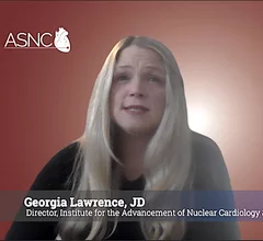
Georgia Lawrence, JD, with ASNC, explains how CMS uses hospital reported data to develop cardiac PET payment rates.

Understanding hospital billing for cardiac PET/CT

Significant fluctuations in PET and CT reimbursement rates have made it especially challenging to keep up with this complex topic. We spoke to an expert to learn more.

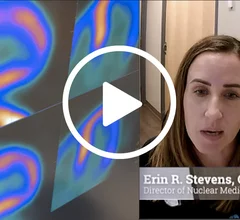
Erin Stevens explained the technical aspects for a successful PET program. #ASNC #thinkPET

Key steps to implementing a successful PET program

Getting technologists, physicians and administrators to work together is a crucial part of any PET program. Research is also essential. Cardiovascular Business spoke to a specialist on the topic to learn more.

Warning: Sidelined reactor could trigger medical isotope shortage

The impending shortage pertains to molybdenum-99, or Mo-99. A structural issue has occurred with a pipe within the high-flux reactor in Petten, Netherlands, which will require an immediate repair. 

Video of Jamshid Maddahi, MD, UCLA, explaining why flurpiridaz will change cardiac imaging and increase the adoption of PET cardiac imaging.

A closer look at how FDA's flurpiridaz approval will impact nuclear cardiology

The newly approved PET radiotracer is expected to improve patient care significantly. “We have been able to reach the pinnacle of myocardial perfusion imaging with flurpiridaz," one expert said.

Banner ASC in Sun City, Arizona.

Low doses of radiation still increase risk of leukemia, lymphoma and myeloma

A new study found cumulative radiation exposure of 16 mSv to red bone marrow can cause an increased rate of some hematological malignancies.

AURORA trial results for Flurpiridaz F-18 PET vs. SPECT discussed by principal investigator Jamshid Maddahi, MD.

'This could be a paradigm shift': How PET with newly approved flurpiridaz compares to SPECT

GE HealthCare's flurpiridaz, the PET radiotracer that recently received FDA approval, offers several key benefits over SPECT. Jamshid Maddahi, MD, discussed the details in an exclusive interview. 

Around the web

Ron Blankstein, MD, professor of radiology, Harvard Medical School, explains the use of artificial intelligence to detect heart disease in non-cardiac CT exams.

Eleven medical societies have signed on to a consensus statement aimed at standardizing imaging for suspected cardiovascular infections.

Trimed Popup
Trimed Popup