Nuclear Medicine

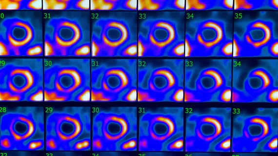
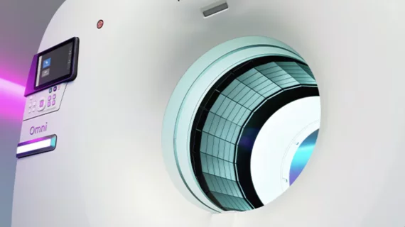
Nuclear medicine (also called molecular imaging) includes positron emission computed tomography (PET) and single photon emission computed tomography (SPECT) imaging. Nuclear imaging is achieved by injecting small amounts of radioactive material (radiopharmaceuticals) into patients before or during their scan. These can use sugars or chemical traits to bond to specific cells. The radioactive material is taken up by cells that consume the sugars. The radiation emitted from inside the body is detected by photon detectors outside the body. Computers take the data to assemble images of the radiation emissions. Nuclear images may appear fuzzy or ghostly rather than the sharper resolution from MRI and CT.  But, it provides metabolic information at a cellular level, showing if there are defects in the function of the heart, areas of very high metabolic activity associated with cancer cells, or areas of inflammation, data not available from other modalities. These noninvasive imaging exams are used to diagnose cancer, heart disease, Alzheimer’s and Parkinson’s disease, bone disorders and other disorders. 
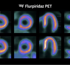
AURORA trial results for Flurpiridaz F-18 PET vs. SPECT discussed by principal investigator Jamshid Maddahi, MD.

'This could be a paradigm shift': How PET with newly approved flurpiridaz compares to SPECT

GE HealthCare's flurpiridaz, the PET radiotracer that recently received FDA approval, offers several key benefits over SPECT. Jamshid Maddahi, MD, discussed the details in an exclusive interview. 

The new cardiac PET radiotracer flurpiridaz F-18 is posed to be a major game changer and will likely lead to increased adoption of cardiac PET.

FDA approves GE HealthCare's flurpiridaz F-18 PET radiotracer for CAD

The newly approved radiotracer is seen as a major step forward for nuclear imaging technology. Specialists have been looking forward to its arrival on the market for quite some time.

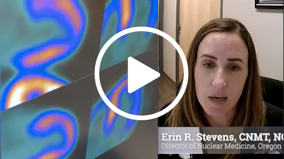
Erin R. Stevens, CNMT, NCT, director of nuclear medicine at Oregon Heart Center, said training physicians for what is needed in cardiac PET documentation is key for preventing issues with prior authorizations. ASNC photo

Good documentation is the key to cardiac PET prior authorizations

Knowing what, exactly, is needed in cardiac PET documentation is one of the best ways to limit prior authorization issues.

PHOTO GALLERY: ASNC2024, the American Society of Nuclear Cardiology's annual meeting

The three-day event attracted nuclear cardiology specialists from all over the world.

Numerous advances in cardiac nuclear imaging led American Society of Nuclear Cardiology (ASNC) President-elect Panithaya Chareonthaitawee, MD, to predict "We are on the edge of a new journey in nuclear cardiology, and the opportunities before us are just as vast as they are exciting and promising."

ASNC president-elect predicts unprecedented innovation in nuclear cardiology

"We are on the edge of a new journey in nuclear cardiology," explained ASNC President-elect Panithaya Chareonthaitawee, MD.


doctor examines patient data on their tablet

FDA sees potential in new PET imaging agent for cardiac amyloidosis

Early evidence suggests a new PET imaging agent from California-based Attralus can help evaluate all varieties of systemic cardiac amyloidosis. It has now been granted the FDA's breakthrough therapy designation. 

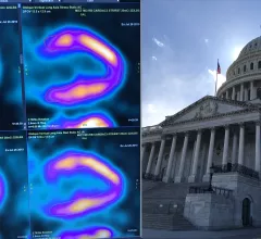
The American Society of Nuclear Cardiology (ASNC) is asking Congress to repeal the appropriate use software provision mandate, which physicians say is an obstacle to efficient care.

ASNC asks Congress to officially repeal the AUC mandate for advanced medical imaging

Medicare rescinded the provision in the 2024 Medicare Physician Fee Schedule, but the law remains on the books. 

How the proposed Medicare Physician Fee Schedule could impact nuclear cardiology

The American Society of Nuclear Cardiology explored some key points included in the 2025 Medicare Physician Fee Schedule proposed rule.

Around the web

Several key trends were evident at the Radiological Society of North America 2024 meeting, including new CT and MR technology and evolving adoption of artificial intelligence.

Ron Blankstein, MD, professor of radiology, Harvard Medical School, explains the use of artificial intelligence to detect heart disease in non-cardiac CT exams.