
Extracellular volume assessments can make cardiac CT more valuable than ever
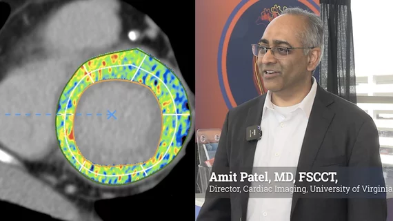
Extracellular volume (ECV) assessment is emerging as a way for cardiac CT to move beyond its traditional focus on the coronary arteries, offering clinicians a noninvasive method to characterize myocardial tissue and identify conditions such as cardiac amyloidosis, aortic stenosis, diffuse fibrosis and edema.
Amit Patel, MD, director of cardiac imaging and a professor of medicine at the University of Virginia Health System in Charlottesville, spoke to Cardiovascular Business about the growing role of ECV assessment at SCCT2026, the Society of Cardiovascular Computed Tomography (SCCT) annual meeting.
Although ECV measurement by CT was developed around the same time as the technique used in cardiac MRI, Patel said MRI has historically made greater use of the technology because of its emphasis on heart failure and myocardial disease. Cardiac CT, by comparison, has largely been used to assess coronary arteries. But that distinction is beginning to change as CT technology and clinical applications expand.
“CT offers a wealth of data,” Patel said. “For most of its history, we've been very focused appropriately so on the coronary arteries, but all along, you've had the whole heart there.”
CT looking beyond the coronary arteries
ECV refers to the space outside of the myocardial cells, including the interstitial tissue between cells and around blood vessels. Under normal circumstances, that space contains relatively small amounts of fibrotic and other interstitial material.
Disease can expand the extracellular space. Fibrosis is known to accumulate there, while conditions such as amyloidosis can cause abnormal proteins to be deposited. Inflammation and edema can also increase the amount of extracellular fluid.
CT can quantify this space by using an iodine-based contrast agent. After contrast is injected, iodine moves from the blood vessels into the extracellular space. By acquiring images after contrast has reached equilibrium and comparing the myocardial and blood-pool measurements, clinicians can estimate the proportion of myocardial tissue occupied by the extracellular space.
He said this is the same basic principle used with gadolinium contrast in MRI ECV assessments.
A potential opportunity in aortic stenosis and amyloidosis
One of the most important potential applications of CT-derived ECV is identifying cardiac amyloidosis in patients undergoing evaluations for severe aortic stenosis. The American Heart Association (AHA) has made this a targeted area for better assessments, because aortic stenosis is still underdiagnosed and undertreated.
Patel said an ECV of roughly 30% or less is generally considered normal, while substantially higher values—particularly measurements in the 40% to 60% range—can raise suspicion for amyloid infiltration.
The overlap between aortic stenosis and amyloidosis is particularly relevant because patients undergoing transcatheter aortic valve replacement (TAVR) already routinely undergo cardiac CT as part of their procedural evaluation, and this could be an easy add-on assessment.
Historically, physicians may have been less likely to consider amyloidosis in a patient whose thickened heart muscle could be explained by aortic stenosis. Increasing evidence suggests the two conditions can coexist. That makes existing CT examinations a potential opportunity to identify an otherwise unrecognized disease.
“If you diagnose it, then you have the opportunity to really change someone's outcome by getting them on the right treatments,” Patel said.
The issue has become more consequential as drug treatments for cardiac amyloidosis have expanded. Patel noted that the disease, once considered rare, is increasingly recognized as more common in older adults, particularly men. He said available therapies can improve heart failure symptoms and quality of life and increase survival in some patients.
ECV remains underused in CT
Despite its potential, ECV assessment is not yet routinely incorporated into cardiac CT practice. Patel attributed part of the problem to workflow. Centers need software capable of performing the measurements efficiently, and acquiring additional delayed images can increase scan time and radiation exposure.
Still, he said the potential diagnostic benefit may justify the additional imaging, particularly in patients with severe aortic stenosis.
“If you had to do it manually, it's not the hardest thing in the world to do,” Patel said, adding that it may add several minutes to the examination.
He also sees a potential role for delayed CT imaging in patients presenting with chest pain and mildly elevated troponin levels when coronary CT does not explain the cause. Myocarditis or another myocardial process could potentially be detected through this tissue characterization.
However, Patel cautioned that the evidence is less established in this younger patient population and the implications for treatment are not yet as clear.
CT and MRI moving closer together
The expanding use of ECV is part of a broader evolution in cardiac CT, Patel explained.
Modern CT can provide information about cardiac anatomy, ventricular function, valves and myocardial tissue in addition to coronary arteries. Advances in radiation-dose reduction and newer technologies, including photon-counting CT, are further expanding the potential capabilities of the modality.
For decades, cardiac MRI has been regarded as the comprehensive modality for myocardial assessment, while CT has held a distinct advantage for coronary imaging. Patel suggested that technological advances could increasingly blur that distinction.
“We used to describe MRI as the one-stop shop,” he said. “CT is really great at the coronary arteries and now we're going to start to see that, hey, maybe it can get the function in the myocardial tissue characterization and myocardial scarring and really, maybe that's the modality that gives us everything we want.”
Whether CT ultimately reaches that point remains to be seen. But the increasing ability to extract myocardial information from scans already being performed could give cardiac CT a broader role in evaluating patients with heart failure and structural heart disease. CT is also much more widely available than cardiac MR.

Dave Fornell has covered healthcare for more than 17 years, with a focus in cardiology and radiology. Fornell is a 5-time winner of a Jesse H. Neal Award, the most prestigious editorial honors in the field of specialized journalism. The wins included best technical content, best use of social media and best COVID-19 coverage. Fornell was also a three-time Neal finalist for best range of work by a single author. He produces more than 100 editorial videos each year, most of them interviews with key opinion leaders in medicine. He also writes technical articles, covers key trends, conducts video hospital site visits, and is very involved with social media. E-mail: [email protected]