Magnetic Resonance Imaging

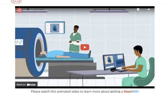
Cardiac magnetic resonance imaging MRI is used as both a functional and anatomical cardiac imaging test. It offers excellent soft tissue detail and the ability to quantify cardiac function. MRI scans can be performed with or without gadolinium contrast depending on what information is needed. Unlike computed tomography (CT), MRI does not use X-ray radiation, but patients with metal implants may have contraindications for MRI use because MR will heat up most metal objects. MRI exams usually take much longer than CT scans. How does MRI work? MR creates images by using powerful magnets to polarize hydrogen atoms in water (the body is made of of more than 80% water) so they face in one direction. A radiofrequency pulse is then used to ping these atoms, causing them to wobble, or resonate. The MRI coils detect this and computers can assemble images from the signals. Basic MRI scans will focus on the resonance of fat and water in two different sequences, which highlight and contrast different features in the anatomy.
Cardiology now has more than 100 FDA cleared AI algorithms

Cardiology makes up 10% of the 692 market-cleared clinical AI algorithms in the FDA’s latest update on the number of patient-facing AI now commercialized in the U.S. Experts share their thoughts on how it is being used.

artificial intelligence consultation

Deep learning in cardiovascular imaging: 4 key takeaways for cardiologists

More and more AI algorithms are being trained to learn, think and act like a human physician. What does this mean for the future of cardiovascular imaging as time goes on?

Thumbnail

Multidisciplinary quality improvement initiative significantly improves on-time starts for cardiac MRI

Such delays can result in longer patient fasts, extended wait times, and poor synchronization of anesthesia induction, researchers detailed in JACR

Thumbnail

AFib recurrence during the 90-day blanking period after ablation—nothing serious or a sign of trouble?

Asking patients to use smartphone-powered ECG devices following catheter ablation procedures could be incredibly beneficial, according to new findings published in JACC: Clinical Electrophysiology

HCM expert Sherif F. Nagueh, MD, FACC, FASE, professor of cardiology, Methodist DeBakey Heart and Vascular Center, explains the key points of HCM imaging. #ASE #HCM

How medical imaging helps cardiologists treat hypertrophic cardiomyopathy

Hypertrophic cardiomyopathy expert Sherif F. Nagueh, MD, examined imaging's role in treating this serious condition. 

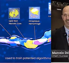
Marcelo DiCarli, MD, chief, division of nuclear medicine and molecular imaging, executive director for the cardiovascular imaging program, Brigham and Women's Hospital, discusses how artificial intelligence (AI) is impacting cardiac imaging.

What is the ROI for adopting AI in cardiac imaging?

Marcelo DiCarli, MD, and Rob Beanlands, MD, discussed the long-term value of investing in the development and implementation of AI technologies. 

Janet Wei at Cedars-Sinai Hospital explains INOCA and MINOCA at ACC 2023.

Understanding INOCA and MINOCA epidemiology

Janet Wei, MD, associate medical director of the Biomedical Imaging Research Institute, and co-director of the stress echocardiography lab at Cedars-Sinai Hospital, explains the current information on the INOCA and MINOCA.

Advanced imaging does not always improve outcomes, data suggest

Although advanced imaging exams have proven benefits in defining disease severity, new data indicate that more sophisticated studies might not impact outcomes as much as previously thought. 

Around the web

Ron Blankstein, MD, professor of radiology, Harvard Medical School, explains the use of artificial intelligence to detect heart disease in non-cardiac CT exams.

Eleven medical societies have signed on to a consensus statement aimed at standardizing imaging for suspected cardiovascular infections.

Trimed Popup
Trimed Popup