Magnetic Resonance Imaging

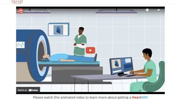
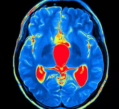
Cardiac magnetic resonance imaging MRI is used as both a functional and anatomical cardiac imaging test. It offers excellent soft tissue detail and the ability to quantify cardiac function. MRI scans can be performed with or without gadolinium contrast depending on what information is needed. Unlike computed tomography (CT), MRI does not use X-ray radiation, but patients with metal implants may have contraindications for MRI use because MR will heat up most metal objects. MRI exams usually take much longer than CT scans. How does MRI work? MR creates images by using powerful magnets to polarize hydrogen atoms in water (the body is made of of more than 80% water) so they face in one direction. A radiofrequency pulse is then used to ping these atoms, causing them to wobble, or resonate. The MRI coils detect this and computers can assemble images from the signals. Basic MRI scans will focus on the resonance of fat and water in two different sequences, which highlight and contrast different features in the anatomy.
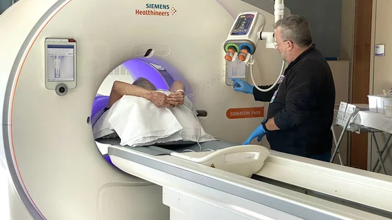

Patients with non-MR compatible cardiac devices, including pacemakers and ICDs, can safely undergo MRI exams

"We had no issues with any of the patients and no harm to the devices," one researcher reported. The analysis included more than 500 patients. 

Thumbnail

Brain MR imaging linked to better outcomes for patients with acute ischemic stroke

Brain MRI scans do lead to increased costs, the authors noted, but the clinical benefits are significant.

Thumbnail

Stress CMR imaging a safe, valuable option when treating HFrEF patients

The study's authors said their findings suggest "an increased value of stress CMR in this population." 

Thumbnail

MRI detects heart failure risk in patients with dilated cardiomyopathy

About 40% of individuals had scarring patterns on their heart muscle visible during imaging, which was associated with a greater risk of suffering a major cardiac event.

Thumbnail

Cardiac MR comparable to FFR for evaluating nonculprit lesions after STEMI

A study published in the latest edition of JACC: Cardiovascular Imaging suggests cardiac magnetic resonance imaging can be as effective as measurements of fractional flow reserve in evaluating nonculprit lesions after STEMI.

The economic impact of MR on heart failure patients

Newly diagnosed HF patients with concomitant mitral regurgitation can expect more admissions, longer hospital stays and pricier medical bills than HF patients without MR, according to an analysis published in the American Journal of Cardiology.

Thumbnail

Cardiac MRI-derived T2 mapping may help heart failure patients

Researchers used T2 mapping taken from weekly cardiac MRIs to help identify cardiotoxicity at an early stage, according to results of a pig study published Feb. 18 in the Journal of the American College of Cardiology. The findings could help cancer patients at risk of chemo-induced heart failure.

Thumbnail

T2 mapping may uncover cardiotoxic marker early enough to prevent heart failure

T2 mapping derived from weekly cardiac MRIs helped researchers identify cardiotoxicity at an early and reversible stage, a finding which may have implications for cancer patients at risk of chemotherapy-induced heart failure.

Around the web

Ron Blankstein, MD, professor of radiology, Harvard Medical School, explains the use of artificial intelligence to detect heart disease in non-cardiac CT exams.

Eleven medical societies have signed on to a consensus statement aimed at standardizing imaging for suspected cardiovascular infections.

Trimed Popup
Trimed Popup