Magnetic Resonance Imaging

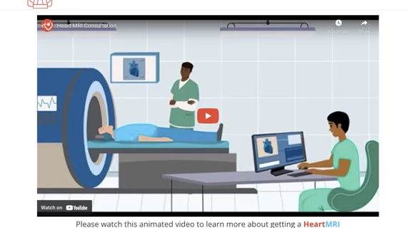
Cardiac magnetic resonance imaging MRI is used as both a functional and anatomical cardiac imaging test. It offers excellent soft tissue detail and the ability to quantify cardiac function. MRI scans can be performed with or without gadolinium contrast depending on what information is needed. Unlike computed tomography (CT), MRI does not use X-ray radiation, but patients with metal implants may have contraindications for MRI use because MR will heat up most metal objects. MRI exams usually take much longer than CT scans. How does MRI work? MR creates images by using powerful magnets to polarize hydrogen atoms in water (the body is made of of more than 80% water) so they face in one direction. A radiofrequency pulse is then used to ping these atoms, causing them to wobble, or resonate. The MRI coils detect this and computers can assemble images from the signals. Basic MRI scans will focus on the resonance of fat and water in two different sequences, which highlight and contrast different features in the anatomy.

TVT2017: 4C MR therapy could be safer than other TMVR approaches

A new mitral regurgitation (MR) therapy designed to treat patients with structural heart disease was shown to be a safer treatment option than other transcatheter mitral valve replacement (TMVR) in a presentation at this year’s Transcatheter Valve Therapies (TVT) conference in Chicago.

FDA approves Arterys cardiac MR imaging software

Cloud-based medical imaging software company Arterys announced Wednesday that it has received 510(k) clearance from the FDA for its Arterys Software. The product provides physicians with quick cardiac MR images.

Providers outside the U.S. consider Cerner more than any other EMR vendor

In a newly published KLAS report entitled "Global HIT Trends: Which Vendors are Providers Betting On?" KLAS interviewed providers around the world to better understand their EMR purchasing plans.

Thumbnail

On the QT: EMR Drug Alerts That Physicians Value

Patient safety is a major concern for every health professional. However, for cardiologists, the interaction of certain drugs and heart conditions make vigilance against contraindications and complications an added battle. EMRs can help, but only if they work reliably and clinicians observe alerts.

Thumbnail

CMR may help predict stroke risk in patients with AF

Based on cardiac magnetic resonance (CMR) measurements in patients with atrial fibrillation, there was an association between left atrial reservoir dysfunction and a prior history of stroke or transient ischemic attack, according to a retrospective study.

3D CMR shines as diagnostic tool for detecting CAD

Whole-heart 3D myocardial perfusion cardiac MR (CMR) accurately detected coronary artery disease (CAD) in a study that used fractional flow reserve as a reference, making CMR a possible contender to techniques that are invasive and expose patients to ionizing radiation.

Thumbnail

CMR helps hone treatment for recurrent pericarditis

Cardiac MR (CMR) can help guide therapy in patients with recurrent pericarditis. One center reported patients who underwent imaging received a smaller total dose of steroids and experienced fewer recurrences than patients who did not get scans.

Thumbnail

Intra-arterial therapy shines in MR CLEAN stroke trial

Giving patients with certain acute ischemic strokes intra-arterial treatment in addition to tissue plasminogen activator led to better outcomes compared with usual care, MR CLEAN researchers reported online Dec. 17 in the New England Journal of Medicine.

Around the web

Ron Blankstein, MD, professor of radiology, Harvard Medical School, explains the use of artificial intelligence to detect heart disease in non-cardiac CT exams.

Eleven medical societies have signed on to a consensus statement aimed at standardizing imaging for suspected cardiovascular infections.

Trimed Popup
Trimed Popup