
Seven-second X-ray assesses severity of regurgitation congenital heart defect patients
A low-dose, moving X-ray exam may be able to expand access to patients to evaluate the severity of pulmonary valve regurgitation following surgical repair of the congenital heart defect Tetralogy of Fallot, according to a study published in the journal Radiology.[1]
Pulmonary valve regurgitation is a significant complication of these surgeries and there is often a gap in care between basic echocardiography and advanced cardiac MRI. Researchers at Kyushu University in Japan looked at using dynamic, moving X-rays to expanding diagnostic options when cardiac MRI is unavailable or contraindicated. These exams can be performed on standard and X-ray systems that are dynamic digital radiography (DDR) capable, such as those produced by Shmiadzu and Konica-Minolta.
Researchers said the new technique only requires a scan of 7 seconds to capture sequential chest images while the patient holds their breath. It was found to have an accuracy rate of 93%. They said this method can be used to help diagnose patients who cannot undergo traditional diagnostic methods, it can improve access to accurate diagnosis, and offers greatly improve efficiency.
Tetralogy of Fallot (TOF) is the most common cyanotic congenital heart defect that affects about 1 in 3,500 newborns. The condition usually requires multiple surgeries and imaging followups.
“Thankfully, with advances in surgical techniques and patient care, today over 90% of patients survive into adulthood,” explained study first author and Assistant Professor Yuzo Yamasaki of Kyushu University Hospital Radiology Center, in a statement. “However, pulmonary regurgitation (PR) is a common long-term complication after surgical repair that can lead to increased risk of sudden cardiac arrest if left untreated. Monitoring PR severity is essential to determine when patients should be treated.”
While cardiac MRI is the standard of care for quantifying the severity of PR. The procedure can be limiting to patients since MRIs can be expensive and require expertise and equipment typically available at specialized facilities. Additionally, MRIs cannot be performed in patients who are contraindicated, such as those with incompatible pacemakers or defibrillators, or who are claustrophobic.
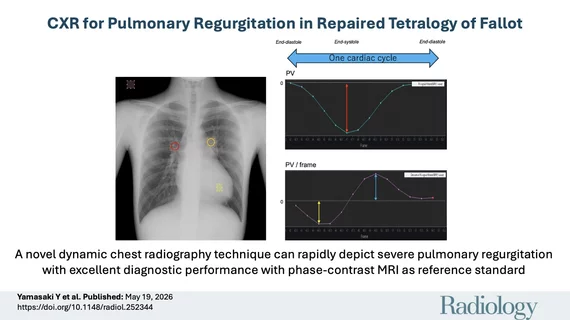
“Dynamic cardiac radiography (DCR) images are usually assessed visually. However, for this study, we analyzed temporal changes in pixel values over the pulmonary arteries in captured sequential images. These changes were converted into waveforms, allowing us to quantify blood flow dynamics,” Yamasaki explained. “In patients with PR, blood flows back into the right ventricle during each heartbeat. The more severe the regurgitation, the more blood flows and that shows up as a more prominent waveform."
Researchers tested this new technique in 58 post-surgical patients and 14 healthy volunteers.
Unlike MRI or CT, the exam does not require contrast. It also has a minimal radiation does of about 0.2 mSv, which is far lower than a standard chest CT of 6 mSv.
The authors said implementation of their new technique may broaden access and reduce healthcare costs.
Yamasaki said DCR might also be able to diagnose other heart diseases, like heart failure and pulmonary hypertension
“We are currently planning a multicenter study to further validate these findings and hopefully establish DCR in routine clinical practices,” he said.

Dave Fornell has covered healthcare for more than 17 years, with a focus in cardiology and radiology. Fornell is a 5-time winner of a Jesse H. Neal Award, the most prestigious editorial honors in the field of specialized journalism. The wins included best technical content, best use of social media and best COVID-19 coverage. Fornell was also a three-time Neal finalist for best range of work by a single author. He produces more than 100 editorial videos each year, most of them interviews with key opinion leaders in medicine. He also writes technical articles, covers key trends, conducts video hospital site visits, and is very involved with social media. E-mail: [email protected]