
New guideline standardizes echo guidance for mitral transcatheter edge-to-edge repairs
The American Society of Echocardiography (ASE) has released a new guideline standardizing intraprocedural imaging for patients undergoing mitral valve transcatheter edge-to-edge repair (M-TEER).[1]
M-TEER is a rapidly expanding minimally invasive, transcatheter valve leaflet clipping procedure that decreases or eliminates mitral regurgitation. However, these procedures have moved outside of structural heart centers of excellence and are now performed by operators and interventional echocardiographers with varying levels of imaging experience and expertise. The new guideline is designed to provide these new users with comprehensive recommendations on best practices for image acquisition, communication and procedural guidance to promote consistency and improved outcomes. ASE said clinicians doing these procedures must have a detailed understanding of mitral valve anatomy, the ability to manipulate two-dimensional and three-dimensional images in real time, and enough procedural experience to understand problems that may be seen on imaging during procedures.
“Until now, there was not a unified, contemporary framework for intraprocedural imaging during M-TEER procedures,” said ASE Past President and Guideline Chair Stephen Little, MD, FRCPC, FACC, FASE, an interventional cardiologist and medical director of the valve clinic at the Houston Methodist DeBakey Heart and Vascular Center, in a statement. “This guideline establishes a standardized approach to imaging and communication among interventional echocardiographers and interventional cardiology or surgical operators to reduce variability and enhance procedural success.”

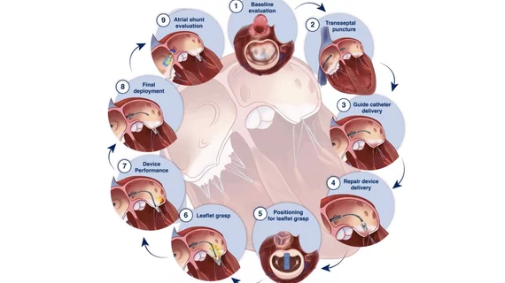
ASE M-TEER guidelines image showing live 3D MPR echo can be used to guide clipping devices into the proper alignment with the mitral valve leaflets for optimal grasping.
Procedural imaging can be performed with 2D transesophageal echocardiography (TEE), but the guideline writing group emphasized the importance of more contemporary 3D multiplanar reconstruction (3D MPR) for M-TEER interventions because the technology increases the precision of device implantation. Clear, 3D imaging and simultaneous multiplanar views of the valve offers a much better understanding of the leaflets and orientation of the clipping device in order to properly grasp the leaflets for optimal outcomes.
“3D MPR has become an essential tool for guiding M-TEER procedures with greater precision,” said ASE Board of Directors member and Guideline Co-Chair Nishath Quader, MD, professor of medicine in the cardiovascular division at Washington University School of Medicine in St. Louis, in the same statement. “We hope this guideline encourages its consistent use among imaging specialists involved in these procedures to optimize patient outcomes.”
Read the complete “Guidelines for the Intraprocedural Imaging for M-TEER: Recommendations from the American Society of Echocardiography.”

ASE M-TEER guidelines image showing how live 3D MPR echo imaging can optimize the transseptal puncture, where location is critical to enable the correct catheter geometry positioing to grasp the leaflets.

Dave Fornell has covered healthcare for more than 17 years, with a focus in cardiology and radiology. Fornell is a 5-time winner of a Jesse H. Neal Award, the most prestigious editorial honors in the field of specialized journalism. The wins included best technical content, best use of social media and best COVID-19 coverage. Fornell was also a three-time Neal finalist for best range of work by a single author. He produces more than 100 editorial videos each year, most of them interviews with key opinion leaders in medicine. He also writes technical articles, covers key trends, conducts video hospital site visits, and is very involved with social media. E-mail: [email protected]