Cardiac Imaging

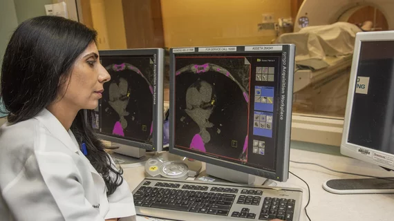
While cardiac ultrasound is the widely used imaging modality for heart assessments, computed tomography (CT), magnetic resonance imaging (MRI) and nuclear imaging are also used and are often complimentary, each offering specific details about the heart other modalities cannot. For this reason the clinical question being asked often determines the imaging test that will be used.

Thumbnail

Document offers guidance on curbing radiation exposure during CV imaging

An expert consensus document published May 2 in the Journal of the American College of Cardiology offers recommendations for limiting radiation exposure to both patients and medical personnel during cardiovascular imaging procedures.

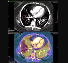
Example of spectral, or dual-energy CT, confirming a pulmonary embolism (PE). Image courtesy of Philips Healthcare

MRA vs CTA for evaluating pulmonary embolism: Does the chosen modality impact downstream imaging utilization?

MR angiography (MRA) is a relatively new alternative to CT angiography (CTA) for the evaluation of suspected pulmonary embolism (PE) and it even has one major advantage over CTA: it does not expose patients to ionizing radiation.

Thumbnail

Machine learning in cardiac CT could be useful—if clinicians put aside their skepticism

Machine learning in cardiovascular CT has the potential to improve disease diagnosis and predictive models, according to the authors of a new paper, but limitations with its applications still remain.

3D brain imaging technology earns FDA approval

An imaging technology called RAPID CTA that enables 3D visualization of a brain’s blood vessels has received FDA clearance, iSchemaView announced May 1.

Thumbnail

Novel device treats pulmonary embolism without clot-dissolving drugs

A new device allows clinicians to remove blood clots from the pulmonary arteries and improve right ventricular function without the use of thrombolytics, according to a late-breaking clinical study presented April 26 at SCAI 2018 in San Diego.

Thumbnail

James K. Min named editor-in-chief of Journal of Cardiovascular Computed Tomography

James K. Min, MD, has been chosen to be the new editor-in-chief of the Journal of Cardiovascular Computed Tomography (JCCT), the official journal of the Society of Cardiovascular Computed Tomography (SCCT). He will serve a five-year term.

James Min named editor-in-chief of Journal of Cardiovascular Computed Tomography

Vienna, VA (April 25, 2017) — James K. Min, MD, FSCCT, has been selected for a five-year term as the new editor-in-chief of the Journal of Cardiovascular Computed Tomography (JCCT). He in turn has named Todd C. Villines, MD, FSCCT, as executive editor along with Gudrun Feuchtner, MD.

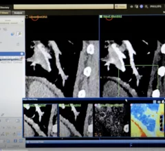
Example of pulmonary embolism (PE) detection on CT and confirmed using spectral CT. Image courtesy of Philips Healthcare

Researchers 'concerned' CT angiography topped MRA in follow-ups for pulmonary embolism

A recent study found whether a patient received MR angiography (MRA) instead of CT angiography (CTA) for a pulmonary embolism (PE) had little effect on the rate of follow-up chest CT or MRI one year later.

Around the web

Several key trends were evident at the Radiological Society of North America 2024 meeting, including new CT and MR technology and evolving adoption of artificial intelligence.

Ron Blankstein, MD, professor of radiology, Harvard Medical School, explains the use of artificial intelligence to detect heart disease in non-cardiac CT exams.