Cardiac Imaging

While cardiac ultrasound is the widely used imaging modality for heart assessments, computed tomography (CT), magnetic resonance imaging (MRI) and nuclear imaging are also used and are often complimentary, each offering specific details about the heart other modalities cannot. For this reason the clinical question being asked often determines the imaging test that will be used.

Thumbnail

Cardiac MRI, angiography produce similar downstream event rates

Patients with stable coronary artery disease (CAD) who were evaluated with either cardiac magnetic resonance (CMR) or conventional angiography showed similar rates of myocardial infarction and death at one and three years of follow-up, according to a study published in JACC: Cardiovascular Imaging.

Thumbnail

Hybrid imaging predicts adverse events for patients with CAD

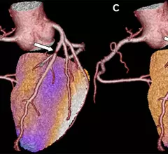
Hybrid imaging with coronary CT angiography (CCTA) and single photon emission CT (SPECT) can provide powerful prognostic value for patients being evaluated for coronary artery disease, according to a single-center study published online July 3 in Radiology.

Thumbnail

Multiple specialties must collaborate to make MRI accessible for all CIED patients

Two neurologists from Stanford University believe their specialty must take the lead on ensuring patients with cardiovascular implantable electronic devices (CIEDs) have access to MRI when necessary.

Thumbnail

Noninvasive HeartFlow test gains commercial coverage from UHC

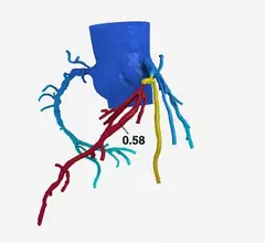
UnitedHealthcare (UHC) will now cover the HeartFlow FFRct Analysis, a noninvasive technology that allows clinicians to view a simulated, three-dimensional model of a patient’s coronary blood flow.

Thumbnail

New PET tracer could boost detection of bacterial heart infections

A novel positron emission tomography (PET) radiotracer could improve upon current methods for identifying bacterial heart infections, according to a presentation at the annual meeting of the Society of Nuclear Medicine and Molecular Imaging in Philadelphia.

Thumbnail

FDA-approved AI echocardiogram software bests cardiologists in reducing LVEF variability

A deep-learning software that can automatically calculate left ventricular ejection fraction (LVEF) with less variability than a cardiologist recently received approval from the U.S. Food and Drug Administration (FDA).

Thumbnail

AI-assisted echocardiogram analysis improves reproducibility of LVEF measurements

A deep-learning algorithm can automatically calculate left ventricular ejection fraction (LVEF) with less variability than cardiologists, according to a study presented June 25 at the American Society of Echocardiography’s annual scientific sessions.
Thumbnail

SNMMI backs updated ASNC guidelines on SPECT MPI

New 2018 guidelines for myocardial perfusion imaging (MPI) protocols on conventional and novel single photon emission computed tomography (SPECT) for nuclear cardiology practitioners has received praise from the Society for Nuclear Medicine and Molecular Imaging (SNMMI).

Around the web

Several key trends were evident at the Radiological Society of North America 2024 meeting, including new CT and MR technology and evolving adoption of artificial intelligence.

Ron Blankstein, MD, professor of radiology, Harvard Medical School, explains the use of artificial intelligence to detect heart disease in non-cardiac CT exams.