Cardiac Imaging

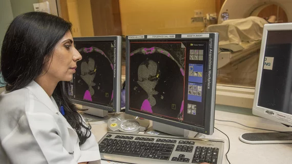
While cardiac ultrasound is the widely used imaging modality for heart assessments, computed tomography (CT), magnetic resonance imaging (MRI) and nuclear imaging are also used and are often complimentary, each offering specific details about the heart other modalities cannot. For this reason the clinical question being asked often determines the imaging test that will be used.

Thumbnail

Noninvasive imaging helps cardiologists map blood flow response to physical stimuli

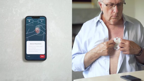
Photoplethysmography—a noninvasive imaging technique that allows clinicians to measure a patient’s pulse wave velocity as blood moves away from their heart—has for the first time linked alterations in the carotid system to changes in physical movement, according to research out of ITMO University in Saint Petersburg, Russia.

Thumbnail

X-ray Vision—Building a Hybrid OR: The Plan, Team & Tactics to Do Imaging Right

Sponsored by Siemens Healthineers

When North Colorado Medical Center in Greeley set out to build a new hybrid OR equipped with robotic angiography, they had no idea the project would set a new bar for project planning and execution across the health system, bring “exponential improvements” in image quality and “exponential reductions” in radiation dose and contrast media, or that they’d finish the project almost a month early without a single change order and $600,000 under budget. Teamwork, meticulous planning and virtual reality-guidance played an essential role in refining and perfecting this image-guided surgery suite even before a pen was put to paper.

Thumbnail

Pre-activating cath labs speeds reperfusion, boosts survival for STEMI patients

ST-segment elevation myocardial infarction (STEMI) patients have better odds of survival if emergency medical services (EMS) personnel alert the hospital where the patient will be arriving ahead of time, according to a study published Sept. 17 in JACC: Cardiovascular Interventions.

SNMMI, ASNC encourage BCBS to expand coverage of cardiac PET

The Society of Nuclear Medicine and Molecular Imaging (SNMMI) and the American Society of Nuclear Cardiology (ASNC) submitted a letter to BlueCross BlueShield (BCBS) of Tennessee urging an expansion of cardiac PET coverage.

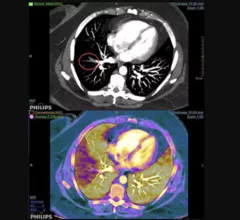
Example of spectral, or dual-energy CT, confirming a pulmonary embolism (PE). Image courtesy of Philips Healthcare

Dual-energy CTA iodine maps offer small benefit in diagnosing pulmonary embolism

Dual-energy (DE) CT iodine maps offer a slight benefit when paired with traditional CT angiography images in diagnosing pulmonary embolism (PE), reported researchers in a Sept. 11 Radiology study.

Body scanners proven safe for patients with ICDs, pacemakers

Millimeter wave body scanners—standard security measures at airports, train stations and public buildings since the 2000s—are completely safe for heart patients with implantable devices, German researchers reported at last month’s ESC Congress.

Thumbnail

New protocol cuts costs, time for CMR imaging

A simplified cardiac magnetic resonance (CMR) protocol using a contrast agent allowed clinicians in Peru to diagnose heart ailments more quickly and cheaply, a new study found. The results, published online Aug. 29 in the Journal of the American Heart Association, highlight a potential pathway to realizing the benefits of CMR in developing nations.

xPerivascular fat attenuation index CT scans from a drug study showing a reduction in coronary wall inflammation. This measure is being tested in trials to see if it can accurately predict which coronary artery lesions will progress to cause heart attacks. The method also can show a reduction in risk with therapy, as seen here.

FAI helps quantify vascular inflammation, predict future CVD events in cardiac imaging patients

The addition of perivascular fat attenuation to routine coronary computed tomography angiography (CTA) improves risk stratification in heart patients, according to research presented at the European Society of Cardiology (ESC)’s annual symposium in Munich, surpassing current prognostic models to re-classify and predict cardiac deaths with more accuracy.

Around the web

Several key trends were evident at the Radiological Society of North America 2024 meeting, including new CT and MR technology and evolving adoption of artificial intelligence.

Ron Blankstein, MD, professor of radiology, Harvard Medical School, explains the use of artificial intelligence to detect heart disease in non-cardiac CT exams.