Cardiac Imaging

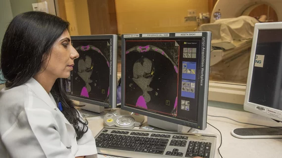
While cardiac ultrasound is the widely used imaging modality for heart assessments, computed tomography (CT), magnetic resonance imaging (MRI) and nuclear imaging are also used and are often complimentary, each offering specific details about the heart other modalities cannot. For this reason the clinical question being asked often determines the imaging test that will be used.

Thumbnail

Prior cardiotoxicity linked to 30% increased risk of CHF during pregnancy

Women with a history of cardiotoxicity from previous cancer treatments are around 30 percent more likely to experience clinical congestive heart failure (CHF) before, during or after pregnancy, according to research published ahead of print in the Journal of the American College of Cardiology.

Thumbnail

Carotid artery MRI boosts risk assessment for CVD, stroke

MRI measurements of carotid wall thickness improve predictions of cardiovascular disease risk more than a commonly used ultrasound-based test, according to research published Oct. 9 in Radiology.

Thumbnail

Angiography-derived FFR accurately diagnoses normal, abnormal vessel function

Fractional flow reserve (FFR) can be closely approximated using conventional coronary angiography and propriety software, according to the FAST-FFR study published online in Circulation. The findings offer a potential route for more patients with suspected coronary artery disease to receive functional assessment of lesions without the need for a guidewire or hyperemic agents.

Thumbnail

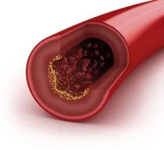
Novel imaging approach tracks atherosclerotic plaque buildup in vivo

Researchers at the University of Tsukuba in Japan have identified a new approach for imaging the progression of atherosclerotic plaque in vivo, according to a study published in Scientific Reports.

Thumbnail

CT-based measurements beat Framingham risk score in predicting future CVD events

Assessment of abdominal aortic calcification using computed tomography (CT) is a highly effective predictor of future heart attack and CVD risk, according to research published in Radiology Oct. 2—more effective even than the Framingham risk score, which has been relied upon for more than two decades.

TCT.18: Test for nonobstructive CAD improves diagnostic accuracy, angina outcomes

With additional testing for nonobstructive coronary artery disease (CAD), researchers effectively diagnosed microvascular and/or vasospastic angina—as well as non-cardiac related chest pain—and used that information to guide therapy decisions and significantly improve angina and quality-of-life outcomes.

TCT.18: COAPT draws superlatives, raises questions about replicating MitraClip’s benefits

Compared to heart failure patients with severe secondary mitral regurgitation who were treated with guideline-directed medical therapy alone, those randomized to a MitraClip procedure plus optimal medical therapy demonstrated relative reductions of 47 percent for heart failure hospitalizations and 38 percent for mortality at two years of follow-up.

Thumbnail

Researchers develop 1st pipeline for automated echocardiogram interpretation

Researchers have developed cardiology’s first pipeline for automated echocardiogram interpretation—an innovation that could cut healthcare costs while expanding care to underserved communities, according to a study published online this week in Circulation.

Around the web

Several key trends were evident at the Radiological Society of North America 2024 meeting, including new CT and MR technology and evolving adoption of artificial intelligence.

Ron Blankstein, MD, professor of radiology, Harvard Medical School, explains the use of artificial intelligence to detect heart disease in non-cardiac CT exams.