Cardiac Imaging

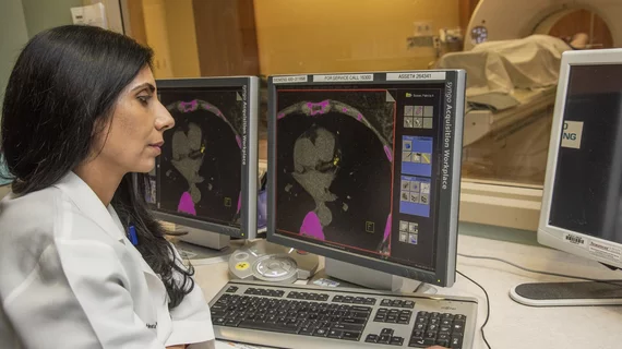
While cardiac ultrasound is the widely used imaging modality for heart assessments, computed tomography (CT), magnetic resonance imaging (MRI) and nuclear imaging are also used and are often complimentary, each offering specific details about the heart other modalities cannot. For this reason the clinical question being asked often determines the imaging test that will be used.

Siemens Healthineers Receives FDA Clearance for TrueFusion Structural Heart Disease Feature

The Food and Drug Administration (FDA) has cleared TrueFusion, a new cardiovascular application from Siemens Healthineers that integrates ultrasound and angiography images to guide cardiac teams when administering treatment for structural heart disease.

ACC releases 10-point criteria for valvular imaging tests

The American College of Cardiology (ACC), partnering with nine other societies, released appropriate use criteria for imaging tests of valvular heart disease. The Sept. 1 document addresses evaluation and use of imaging in diagnosing and managing the condition.

Thumbnail

Value-based Reimbursement Is Here to Stay & Other Lessons from Cath Lab Leadership Boot Camp

The business of cardiology was at the forefront of discussion at the Society for Cardiovascular Angiography and Interventions (SCAI) Cath Lab Leadership Boot Camp in May. Speakers focused on the Medicare Access and CHIP Reauthorization Act of 2015 (MACRA), bundled payment models and value-based reimbursement. Here are a few of the lessons attendees took back to their practices.

Thumbnail

Innovation Competition: At TCT.17, Cardiology’s Disruptors Swim with the Sharks

Cardiology’s Shark Tank will be back for its fourth year when TCT convenes Oct. 29-Nov. 2, in Denver. Program Director Juan F. Granada, MD, shares insights from the conference’s innovation competition.

Thumbnail

Individual feedback key to reducing excessive ECGs

An educational intervention designed to reduce ordering of transthoracic echocardiograms (TTEs) informs physicians about their own test-ordering patterns and how their performance compares to their peers, according to recent research. Such knowledge can reduce inappropriate use of the tests.

ECG reveals clinical findings obscured by left ventricular hypertrophy

A man born with a congenital heart murmur developed systemic arterial hypertension as an adult. At age 50, he went to the hospital because of dyspnea. He also had a pulmonic valve ejection click and murmur of pulmonic stenosis. When at the hospital, his echocardiogram showed hypertrophy of both ventricles as well as an enlarged right atrium.

Thumbnail

Ultrasound may offer nonionizing, contrast-free way to assess coronary flow reserve

To date, clinicians have lacked a noninvasive technique for the assessment of coronary flow reserve, but a new study offers a promise of just such a technique.

Cardiac MRI improves risk stratification for STEMI patients

For revascularized ST-segment elevation myocardial infarction (STEMI) patients, early post-infarction cardiac MRI parameters offer long-term predictive value that is superior to that offered by traditional outcome predictors, a large, long-term European study has found.

Around the web

Several key trends were evident at the Radiological Society of North America 2024 meeting, including new CT and MR technology and evolving adoption of artificial intelligence.

Ron Blankstein, MD, professor of radiology, Harvard Medical School, explains the use of artificial intelligence to detect heart disease in non-cardiac CT exams.