Cardiac Imaging

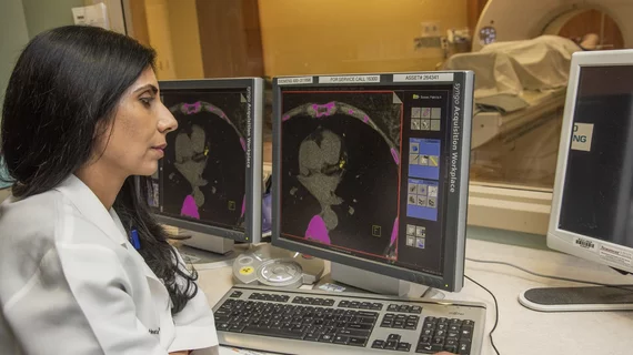
While cardiac ultrasound is the widely used imaging modality for heart assessments, computed tomography (CT), magnetic resonance imaging (MRI) and nuclear imaging are also used and are often complimentary, each offering specific details about the heart other modalities cannot. For this reason the clinical question being asked often determines the imaging test that will be used.

Breast implants could skew ECG results

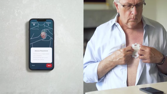
New research from Monaco suggests that breast implants can impede electrocardiogram (ECG) recordings, leading to false readings and potentially incorrect heart attack diagnoses.

TVT2017: 4C MR therapy could be safer than other TMVR approaches

A new mitral regurgitation (MR) therapy designed to treat patients with structural heart disease was shown to be a safer treatment option than other transcatheter mitral valve replacement (TMVR) in a presentation at this year’s Transcatheter Valve Therapies (TVT) conference in Chicago.

Experimental ultrasound tool eradicates blood clots

A new tool that uses low-frequency intravascular ultrasound to break down blood clots that cause deep vein thrombosis is being tested by researchers at North Carolina State University and the University of North Carolina at Chapel Hill.

Thumbnail

New cardiac imaging FFRct gets high rating from BCBS

HeartFlow, a cardiac technology company in Redwood City, California, has received positive reviews on its HeartFlow FFRct, a non-invasive technology that uses coronary CT angiograms to make personalized 3D models of a patient’s arteries.

Hitachi Healthcare Americas to Introduce the “Lisendo 880” at ASE 2017, the New Premium 2D and 3D Cardiovascular Ultrasound System by Hitachi

Premium cardiac and vascular imaging rises to a new standard as Hitachi Healthcare Americas introduces the new premium 2D and 3D cardiovascular ultrasound system, the LISENDO 880, at the American Society of Echocardiography 2017 in Baltimore, Maryland on June 3rd, 2017.

Thumbnail

Imaging study reveals new ways to widen blood vessels

Pharmacological stress agents given to patients during a cardiac stress test can cause severe side effects, but a new study using PET/MR imaging has revealed that using arterial carbon dioxide (PaCO2) can safely and efficiently widen blood vessels.

Thumbnail

Cardiac PET Imaging: When It’s the Right Test at the Right Time

Medical Imaging & Technology Alliance

Diagnosing and treating patients well starts with choosing the most precise test. Cardiac PET imaging is the test of choice more often these days when it comes to assessing coronary artery disease, myocardial perfusion, viability and ventricular function in more complex patients. It makes sense clinically, operationally and financially. It also trumps SPECT in many ways, and can even play a role in cardiac disease prevention.
 

Long-term survivors of pediatric cancer linked to CVD risk

For long-term survivors of pediatric cancer, that early exposure to cardiac radiotherapy may be associated with higher rates of cardiovascular disease (CVD).

Around the web

Several key trends were evident at the Radiological Society of North America 2024 meeting, including new CT and MR technology and evolving adoption of artificial intelligence.

Ron Blankstein, MD, professor of radiology, Harvard Medical School, explains the use of artificial intelligence to detect heart disease in non-cardiac CT exams.