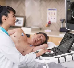
Echocardiography

Cardiac ultrasound uses reflected sound waves (echos) to create images of anatomy inside the body. Echocardiograms are the primary cardiac imaging modality used to assess the heart and diagnose or track cardiac issues. Echo is the gold standard imaging modality to assess the heart, particularly with calculating left ventricular ejection fraction (LVEF), which is a measure of cardiac output. In addition to noninvasive standard transthoracic echo (TTE), invasive transesophgeal echo (TEE) is also used when clearer, more detailed imaging of the heart is needed. Both 3D and 4D echo echo systems are rapidly gaining wider adoption and enable new types of assessments, especially in the structural heart space and in transcatheter procedural guidance. Find news on general ultrasound imaging.
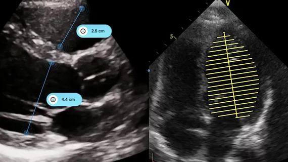
Caption Health gains FDA clearance for AI-powered ejection fraction software

The original software first received FDA clearance back in 2018. This updated version, Caption Health has said, is easier for clinicians to use. 

Thumbnail

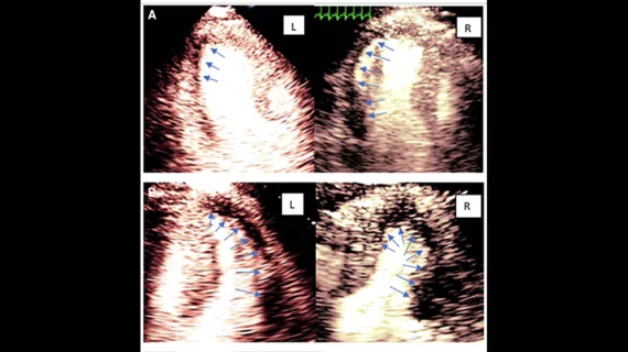
Group publishes new stress echo guidelines for ischemic heart disease

The American Society of Echocardiography released the new recommendations to replace its 2007 edition.

Thumbnail

What’s to Gain from Strain? Experts Say Strain Echocardiography Is on the Path to Acceptance

Despite its diagnostic and prognostic value, speckle-tracking strain echocardiography is underused, some cardiac imagers say. What will it take for adoption to pick up?

Thumbnail

Pocket-sized: Pointers for Using (& Not Using) Handheld Point-of-Care Echocardiography

Handheld POC echo has proven useful in- and outside of the emergency department, but physicians still have reservations.

Thumbnail

Philips updates, expands cardiac ultrasound platform

Royal Philips has rolled out the latest update for its EPIQ CVx and EPIQ CVxi cardiac ultrasound systems, expanding to include automated applications for 2D assessment of the heart and robust 3D measurements of right ventricular volume and ejection fraction.

Thumbnail

CV programs struggling to keep up with growing demand for cardio-oncologists

Cardio-oncology has emerged as an area of rapid growth in the medical community in recent years, owing in large part to an increasing population of cancer survivors.

Thumbnail

Genetic variant linked to chemotherapy-induced cardiomyopathy

The same genetic variants that have been uncovered in patients with two other types of cardiomyopathy are also present in an uncommonly high proportion of people with cancer therapy-induced cardiomyopathy (CCM), researchers reported in Circulation.

Ebit (Esaote Group) and DiA Imaging Analysis have partnered offering Advanced AI-based Cardiac Ultrasound Analysis

BE'ER SHEVA, Israel — Genoa, Italy — DiA Imaging Analysis, a leading provider of artificial intelligence (AI)-powered ultrasound analysis tools, announced today that it has partnered with the Italian healthcare IT company Ebit (Esaote Group), to offer DiA’s LVivo Cardiac Toolbox as an integrated part of Ebit's SUITESTENSA CVIS (Cardiovascular Information System) PACS.

Around the web

Ron Blankstein, MD, professor of radiology, Harvard Medical School, explains the use of artificial intelligence to detect heart disease in non-cardiac CT exams.

Eleven medical societies have signed on to a consensus statement aimed at standardizing imaging for suspected cardiovascular infections.