Echocardiography

Cardiac ultrasound uses reflected sound waves (echos) to create images of anatomy inside the body. Echocardiograms are the primary cardiac imaging modality used to assess the heart and diagnose or track cardiac issues. Echo is the gold standard imaging modality to assess the heart, particularly with calculating left ventricular ejection fraction (LVEF), which is a measure of cardiac output. In addition to noninvasive standard transthoracic echo (TTE), invasive transesophgeal echo (TEE) is also used when clearer, more detailed imaging of the heart is needed. Both 3D and 4D echo echo systems are rapidly gaining wider adoption and enable new types of assessments, especially in the structural heart space and in transcatheter procedural guidance. Find news on general ultrasound imaging.

Digisonics Showcases New Enhancements for Streamlined Cardiovascular Workflows at ASE 2017

HOUSTON (May 30, 2017) – Digisonics (Booth #421) will exhibit its latest functionality for Cardiovascular Information Systems (CVIS) at the American Society of Echocardiography 28th Annual Scientific Sessions in Baltimore, Md.
Thumbnail

Pathways for Quality Care: Echocardiographers Ponder a Playbook for Imaging in Myocardial Infarction

Physicians in fields like cardiology have traditionally looked to clinical practice guidelines to help articulate the best evidence-based care for patients. The rapidly growing movement to value-based care is prompting clinicians—including echocardiographers—to carefully weigh a more focused and integrative approach to delivering consistent, quality medicine: care pathways.

Succeeding with Cancer: Using Imaging to Avoid Treatment-induced Heart Failure

Treating today’s cancer patient no longer means simply targeting the cancer. Given the known cardiotoxicities of some established chemotherapies and the possibility that newer approaches may damage the heart, oncologists, cardiologists and imaging specialists now work together to detect and minimize the risk of treatment-induced heart failure.

Thumbnail

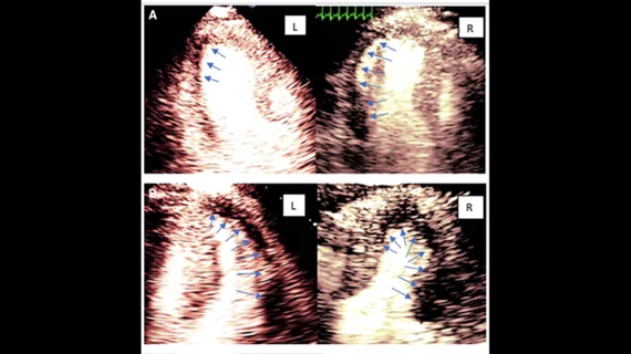
Coding for Clarity: Echocardiography Gains Two New CPT Add-On Codes

The approval of two new Current Procedural Terminology (CPT) codes acknowledges echocardiographic myocardial strain imaging and myocardial contrast perfusion echocardiography as emerging technologies, often a necessary step before a code is promoted to payable status.

Thumbnail

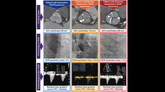
Mitral Mysteries: Imagers Explore a New Frontier in Transcatheter Valve Replacement

Transcatheter aortic valve replacement offers valuable lessons that can be applied to its mitral valve counterpart, according to imagers on the frontlines for both techniques.

Thumbnail

Goal Reversal? Study Suggests Echocardiography Could Have an Underuse Problem

ASE says new data may be a “wake-up call” to recognize the value of echocardiography.  

Thumbnail

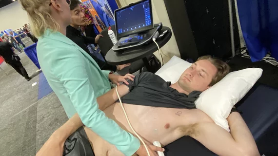
ASE scientific sessions to highlight point-of-care ultrasound, strain imaging and 3D echocardiography

At the American Society of Echocardiography (ASE) annual scientific sessions, the use of point-of-care echocardiography is among the major topics of discussion. The conference takes place from June 12 to June 16 at the Hynes Convention Center in Boston.

Q&A: Contrast for Stress Echo

ASE members discuss recent changes to contrast enhanced stress echo.

Around the web

Ron Blankstein, MD, professor of radiology, Harvard Medical School, explains the use of artificial intelligence to detect heart disease in non-cardiac CT exams.

Eleven medical societies have signed on to a consensus statement aimed at standardizing imaging for suspected cardiovascular infections.

Trimed Popup
Trimed Popup