Echocardiography

Cardiac ultrasound uses reflected sound waves (echos) to create images of anatomy inside the body. Echocardiograms are the primary cardiac imaging modality used to assess the heart and diagnose or track cardiac issues. Echo is the gold standard imaging modality to assess the heart, particularly with calculating left ventricular ejection fraction (LVEF), which is a measure of cardiac output. In addition to noninvasive standard transthoracic echo (TTE), invasive transesophgeal echo (TEE) is also used when clearer, more detailed imaging of the heart is needed. Both 3D and 4D echo echo systems are rapidly gaining wider adoption and enable new types of assessments, especially in the structural heart space and in transcatheter procedural guidance. Find news on general ultrasound imaging.
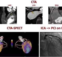
A figure from the 2022 CAD non-invasive imaging guidelines showing a comparison of computed tomography angiography (CTA) and a SPECT-CT vs. an invasive angiogram from the cath lab showing the same blockage in a coronary artery.

New multi-society recommendations highlight role of non-invasive imaging in evaluating coronary artery disease

A new, multi-society document, "Non-Invasive Imaging in Coronary Syndromes," focuses on how multiple imaging techniques can evaluate different aspects of coronary artery disease (CAD), all without the need for invasive angiograms.

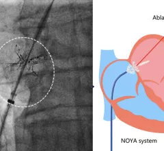
Researchers share first human data on new interventional shunt procedure for HFpEF

The first-in-man study, completed by a team of cardiologists in China, included 10 patients presenting with HFpEF. 

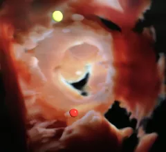
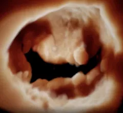
The mitral valve visualized by a a GE NuVision 4D intra-cardiac echo (ICE) catheter a life-like surgical rendering technology on the Vivid E95 cardiac ultrasound system. The catheter was co-developed with Biosense Webster to perform EP procedures. It also can be used in place of TEE in structural heart procedures to eliminate the need for an interventional echocardiography.  #ACC22

Photo Gallery: ACC 2022 in pictures

Click through a wide variety of snapshots from ACC.22 in Washington, D.C. 

Thumbnail

AI distinguishes between a heart attack and takotsubo syndrome more accurately than cardiologists

The advanced AI model outperformed a team of cardiologists, delivering a better AUC and overall accuracy. 

AI models capable of identifying RV, LV dysfunction in ECGs

Specialists used EHR data, imaging results and natural language processing to bring their advanced AI models to life. 

Interventional cardiology procedure in a cath lab at Beaumont Hospital.

PCI boosts survival for ischemic HF patients with moderate-to-severe mitral regurgitation

Mortality rates can be especially high for ischemic HF patients presenting with FMR and LVSD, but PCI may offer some relief. 

William A. Zoghbi, MD, MACC, FAHA, FASE, is the chair of the Department of Cardiology at the Houston Methodist DeBakey Heart and Vascular Center, and past president of both the American College of Cardiology (ACC) and the American Society of Echocardiography (ASE).

VIDEO: New advances in echocardiography

William A. Zoghbi, MD, past president of the ACC and ASE, discussed the latest trends in cardiac ultrasound technology. 

The mitral valve seen on 3D cardiac ultrasound offers a noninvasive surgeon's view of the valve for better diagnostics and pre-planning of structural heart procedures. Example from the GE Vivid E95 FlexiLight photo-realistic 3D/4D lighting system.

Q&A: William Zoghbi breaks down the future of echocardiography

In a new interview, former ACC and ASE President William A. Zoghbi, MD, reviews some of the biggest trends in cardiac ultrasound.

Around the web

Ron Blankstein, MD, professor of radiology, Harvard Medical School, explains the use of artificial intelligence to detect heart disease in non-cardiac CT exams.

Eleven medical societies have signed on to a consensus statement aimed at standardizing imaging for suspected cardiovascular infections.

Trimed Popup
Trimed Popup