Cardiac Imaging

While cardiac ultrasound is the widely used imaging modality for heart assessments, computed tomography (CT), magnetic resonance imaging (MRI) and nuclear imaging are also used and are often complimentary, each offering specific details about the heart other modalities cannot. For this reason the clinical question being asked often determines the imaging test that will be used.

AI cardiology heart artificial intelligence deep learning

FDA approves new AI-powered imaging software from Nanox subsidiary

According to Nanox.AI, previously known as Zebra Medical Vision, the updated software helps "bridge the divide between radiology and cardiology."

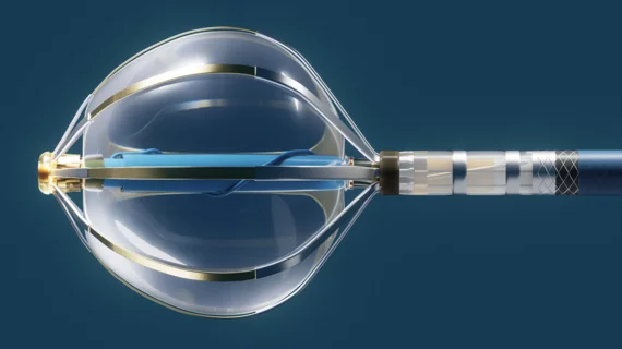
Interventional cardiologists in Canada have performed the world’s first implant of a new coronary sinus reducer designed to treat chest pain patients who see no benefits from other available interventional or surgical treatments. The A-Flux Reducer System was designed and developed by VahatiCor, a new medical device company associated with California-based T45 Labs.

Cardiologists detail world’s first implant of new device for ‘no-option’ chest pain patients

The A-Flux Reducer System by VahatiCor was designed to “conform seamlessly” to any patient’s anatomy, and it can be repositioned or retrieved as necessary.

Ron Blankstein, MD, FACC, FASNC, MSCCT, FASPC, associate director, cardiovascular imaging program, director, cardiac computed tomography, Brigham and Women’s Hospital, and a professor of medicine and radiology, Harvard Medical School, explains why Medicare is proposing increased coronary computed tomography angiography (CCTA) payments to hospitals.

Higher payments for CCTA could bring considerable change to cardiology

Ron Blankstein, MD, explains how and why Medicare is proposing an increase to hospital payments for coronary CT angiography.

Thumbnail

Reduce, reuse, recycle: How cath labs can lower cardiology’s colossal carbon footprint

Cath labs, like radiology suites and operating rooms, are leading contributors to the world’s growing carbon footprint.

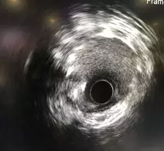
Images coronary calcium imaging by optical coherence tomography (OCT) and high-definition intravascular ultrasound (IVUS).

OCT-guided PCI improves survival after life-threatening heart attacks

Researchers explored data from nearly 4,000 STEMI patients, noting that optical coherence tomography may be a valuable first step of any treatment strategy. 

cardiologists evaluating the human heart to provide a treatment strategy

Managing stable angina: How cardiologists can find the right mix of revascularization, therapy and interventions

The management of stable angina has been evolving at a rapid rate. Using a one-size-fits-all strategy is becoming a thing of the past, replaced by a patient-centered approach that requires open communication and a healthy understanding of recent clinical research.

Ron Blankstein, MD, FACC, FASNC, MSCCT, FASPC, associate director, cardiovascular imaging program, director, cardiac computed tomography, Brigham and Women’s Hospital, and a professor of medicine and radiology, Harvard Medical School, explains a few of the recent advancements in cardiac MRI, nuclear imaging and computed tomography.

Exploring major breakthroughs in cardiac imaging

Ron Blankstein, MD, director of cardiac computed tomography for Brigham and Women’s Hospital, details recent developments in cardiac MRI, nuclear imaging and CT.

The Shockwave Medical M5+ new peripheral intravascular lithotripsy (IVL) catheter cuts procedures times in half and was showed for the first time at the American College of Cardiology (ACC) 2022 meeting. It is designed to break up calcium in heavily calcified arteries to avoid the need for vessel trauma caused by high pressure angioplasty.

Intravascular lithotripsy, the technology at heart of $13B acquisition, linked to positive real-world data

The calcified plaque-shattering technology developed by Shockwave Medical continues to gain momentum. As more care teams start using IVL during PCI, researchers are paying close attention to its long-term impact on patient care.

Around the web

Ron Blankstein, MD, professor of radiology, Harvard Medical School, explains the use of artificial intelligence to detect heart disease in non-cardiac CT exams.

Eleven medical societies have signed on to a consensus statement aimed at standardizing imaging for suspected cardiovascular infections.

Trimed Popup
Trimed Popup