
Cardiologists develop new anchoring technique for challenging TAVR cases
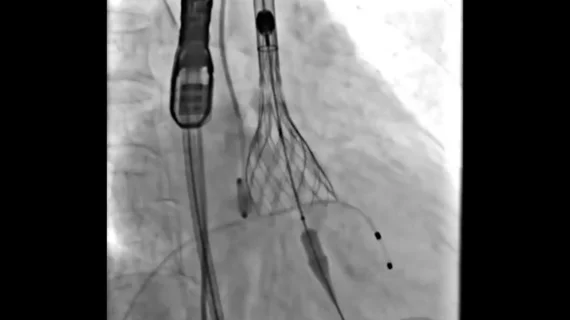
Transcatheter aortic valve replacement (TAVR) can be challenging in patients with pure aortic regurgitation (PAR) and an enlarged left ventricular outflow tract (LVOT), especially when a transcatheter heart valve (THV) with a dedicated anchor is not readily available.
When a team of specialists in China faced that very situation, they developed a modified implant technique—noncoronary sinus pivot implantation (NCPI-plus)—and then wrote about their experience in JACC: Case Reports.[1]
The patient in question was a 74-year-old woman with severe PAR and an enlarged LVOT who could not safely undergo open surgery. The patient presented with resistant hypertension, type 2 diabetes, hyperlipidemia, chronic kidney disease and chronic obstructive pulmonary disease, among other comorbidities. In addition, she had a history of progressive exertional dyspnea, and clinicians identified a diastolic murmur at the left sternal border.
The patient underwent transthoracic echocardiography and CT angiography prior to treatment. Clinicians found that she had a large aortic annulus with no signs of calcification, significant leaflet thickening, or leaflet adhesion. Her left ventricular ejection fraction was 38% and the perimeter of her LVOT was 93.7 mm.
The patient ultimately underwent TAVR with a 30-mm self-expanding valve using the NCPI-plus technique. This approach involves “creating a supra-annular pivot point on the noncoronary sinus side and deep LVOT engagement on the left coronary sinus side, providing a stable anchoring solution.”
The procedure was a success with no signs of paravalvular leakage. The patient was discharged home five days later. Follow-up echocardiography 6 months and then 12 months later demonstrated “excellent valve function, with no measurable transvalvular gradient or aortic regurgitation.”

(A) Post-procedural CT angiography from the present case confirming partial supra-annular anchoring of the transcatheter heart valve frame against the annulus on the noncoronary sinus side (arrow). (B) Three-dimensional reconstruction generated using 3mensio (Pie Medical Imaging) from the patient's original CTA DICOM data, highlighting the anchoring point of the valve frame on the noncoronary sinus side. (C) Illustration of the noncoronary sinus pivot implantation anchoring mechanism. The transcatheter heart valve frame inserts into the muscular tissue on the noncoronary and right-coronary sinus side (arrow), creating a platform that serves as a fulcrum during deployment (reproduced with permission). Images and captions courtesy of Ge et al. and JACC: Case Reports.
Reviewing their experience, the team behind this case highlighted the importance of minimizing the risk of THV migration without introducing additional things that could go wrong. The group also noted that imaging guidance remains a critical part of the TAVR procedure.
“Importantly, this technique does not increase the risk of noncoronary cusp injury, as the pivot point is supported by the robust and dense central fibrous body,” wrote senior author Junbo Ge, MD, director of cardiology at Zhongshan Hospital in Shanghai, China, and colleagues. “Nevertheless, deliberate operator attention (aided by TEE) remains crucial to avoid aggressive manipulation.”
Ge et al. also noted that their team’s previous work with the more traditional NCPI approach was associated with a mean oversizing of 22.1%. For this specific case with the newer technique, on the other hand, oversizing was 16.8%, “suggesting that NCPI-plus may require less oversizing than the standard technique.”
“Current guidelines provide limited specific recommendations for TAVR in PAR patients with enlarged LVOT,” the authors added. “This case demonstrates that the NCPI-plus technique may offer a practical solution, extending the application of the NCPI anchoring principle to this challenging anatomical subset. We recognize that the lack of bench testing is a limitation of this case report, and future studies would be valuable to further validate the anchoring mechanism.”
Click here to read the full breakdown in JACC: Case Reports, an American College of Cardiology journal.

Michael has more than 19 years of experience as a professional writer and editor. He has written at length about cardiology, radiology, artificial intelligence and other key healthcare topics.