Ultrasound

Ultrasound, also referred to as sonography or diagnostic ultrasound, uses high-frequency sound waves to visualize soft tissue. Ultrasounds are frequently ordered to measure fetal anatomy during pregnancy, check for blood clots and to guide needle biopsy procedures of the breast, abdomen and pelvis. The imaging modality does not use any radiation to create images. Find news specific to cardiac ultrasound (echocardiography).

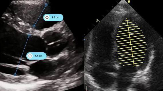
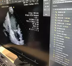
Artificial intelligence automated measurements on an echocardiogram on the Siemens SyngoDynamics cardiovascular imaging and information solution. AI is helping speed workflows and complete tedious tasks faster and more accurately that humans, allowing sonographers and cardiologists to be more efficient. Photo by Dave Fornell

AI technologies to be featured heavily at ASE 2023

Artificial intelligence will be one of the hottest topics at the upcoming American Society of Echocardiography meeting in National Harbor, Maryland. 

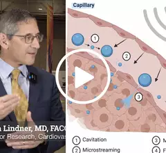
Video of Jon Lindner explaining the use of ultrasound and bubble contrast for therapy.

Cardiac ultrasound could lead to key advances in heart attack care, drug delivery

Jonathan Lindner, MD, offers an update on the use of echocardiography and bubble contrast agents in a therapy role to help revascularize STEMI patients and increase drug and gene delivery.

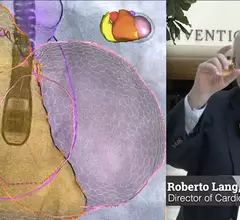
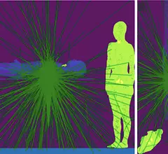
X-ray photon trajectory during the simulation phantom study from the side and top views. Due to scattering of the X-rays when they hit the lower end of the patient bed, exposure in mainly to the lower body of the interventional echocardiographer performing transesophageal echocardiography. The green lines are the scattered photon trajectories calculated by Monte Carlo simulation in the study.

Radiation exposure in the cath lab: Tracking the impact on interventional echocardiographers

Researchers found that echocardiographers in the cath lab are exposed to high doses of radiation on the right half of their body, especially the waist and lower body. 

New ASE guideline outlines training standards for interventional echocardiography

The American Society of Echocardiography (ASE) released a new guideline document that outlines uniform training standards for interventional sonographers guiding structural heart procedures.

artificial intelligence AI deep learning

Cardiologists find AI to be more accurate than sonographers at interpreting echocardiograms

Overall, cardiologists made corrections to 16.8% of AI-generated assessments and 27.2% of sonographer assessments.

Echocardiography expert Patricia A. Pellikka, MD, discussed the trend of increasing artificial intelligence (AI) integration in cardiac ultrasound with Cardiovascular Business at American College of Cardiology (ACC) 2023 meeting.

AI's growing impact on echocardiography

Cardiology has the second largest number of FDA-cleared AI algorithms, and many of them are for cardiac ultrasound. Echocardiography expert Patricia A. Pellikka, MD, discusses this trend and how AI is helping improve echo.

A figure from the ASE pediatric POCUS guidelines showing parasternal short-axis view demonstrating RV dilatation with bowing of the septum into the left ventricle, indicating pulmonary hypertension in this child with shock and pertussis.

New ASE guidance examines cardiac POCUS in children

The recommendations were developed by a writing group comprised of experts from different specialties, offering an array of perspectives and approaches.

caption health GE healthcare echo ultrasound AI

GE Healthcare to acquire AI imaging specialists Caption Health

Caption Health has been a major player in the cardiac imaging space in recent years, gaining attention for its AI-powered echocardiography solutions. 

Around the web

Several key trends were evident at the Radiological Society of North America 2024 meeting, including new CT and MR technology and evolving adoption of artificial intelligence.

Ron Blankstein, MD, professor of radiology, Harvard Medical School, explains the use of artificial intelligence to detect heart disease in non-cardiac CT exams.