
OCT shows potential for image-guided BTK interventions in PAD patients
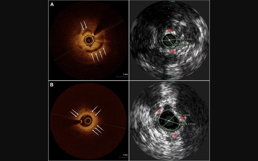
Choosing optical coherence tomography (OCT) over intravascular ultrasound (IVUS) for below-the-knee (BTK) endovascular interventions is a feasible strategy that may offer some key benefits, according to new findings published in JSCAI.[1]
“Durable outcomes in BTK interventions remain challenging—likely owing to several unique aspects of tibial arteries, including smaller diameter vessels with long segment lesions and prevailing circumferential calcium,” wrote first author Jun Li, MD, an interventional cardiologist with the University Hospitals Harrington Heart and Vascular Institute in Cleveland, and colleagues. “Angiographic interpretation of calcium remains subpar compared to intravascular imaging. Although IVUS is reasonable for analysis in the aorto-iliac and femoropopliteal segments, its inability to penetrate through calcium limits its utility in the tibial arteries.”
To learn more about OCT’s potential, Li et al. performed a feasibility study that included data from 44 patients who were enrolled at a single facility from 2021 to 2023. All patients presented with chronic limb-threatening ischemia (CLTI) and underwent angiography, IVUS and OCT.
Overall, measurements captured using OCT images “correlated well” with the IVUS and angiographic analyses. OCT delivered the “most defined” calcium assessment, the group added.
A total of 20 patients had calcium present on the minimum lumen frame of their OCT results. The median calcium arc for these patients was 130° according to OCT and 163° according to IVUS. Median cap thickness was 0.24 mm according to OCT and 0.12 mm according to IVUS.
“In our analysis of IVUS compared to OCT in BTK tibial vessels, significant differences in diameter and area measurements were observed between the two modalities in both preintervention and postintervention cohorts,” the authors wrote. “Nonetheless, correlation testing showed reasonable agreement between the two types of intravascular imaging. The divergence between OCT and IVUS in BTK vessel sizing is consistent with prior coronary studies that have shown discrepancies between these two imaging modalities.”
Meanwhile, median calcium thickness was 0.49 mm according to OCT. IVUS was unable to accurately measure calcium thickness.
Calcium thickness, the authors noted, did appear to potentially be associated with a risk of target lesion revascularization or major amputation. However, it did not appear to influence the risk of all-cause mortality.
Li and colleagues also explained that intravascular imaging may be associated with increased healthcare costs, but those costs have been directly linked to improved patient care. When considering the potential benefits of using these modalities, they wrote, intravascular imaging is a cost-effective treatment strategy when considering the “full episode of care.”
“Further evaluation is needed to assess the utility of OCT to help optimize tibial interventions and improve the durability of revascularization,” the group concluded.
Editorial considers the potential value of OCT
Helena Smith, MD, and Eric A. Secemsky, MSc, MD, shared their perspective on these findings in an editorial that was also published in JSCAI.[2] Smith is a postdoctoral fellow with Beth Israel Deaconess Medical Center (BIDMC) and Secemsky is the director of vascular intervention with BIDMC’s CardioVascular Institute.
The two authors noted that procedural decision making has been slow to change when it comes to the treatment of peripheral artery disease. They also celebrated the work of Li and colleagues as an “important step forward” as cardiologists hope to learn more about how to identify the best treatment strategy for BTK interventions.
In the editorial, Smith and Secemsky pointed to the fact that OCT was linked to significant improvements in terms of visualizing calcific vascular disease.
“Calcium is not simply a marker of disease severity, but it also determines how a lesion mechanically responds to endovascular therapy,” the two co-authors wrote. “Vessel compliance, balloon expansion, acute luminal gain, drug penetration, and, ultimately, procedural durability is influenced by the extent and distribution of calcification. Angiography frequently underestimates calcification, while IVUS, despite its strengths, is limited in its ability to quantify calcium thickness because of acoustic shadowing. OCT overcomes many of these limitations by providing high-resolution visualization of calcium arc, thickness, and longitudinal distribution. In coronary intervention, these features have become central to identifying lesions that require plaque modification before definitive treatment.”
The duo also emphasized that this research is not proof that OCT-guided interventions improve patient outcomes. Instead, the study was focused on feasibility.
“Similarly, the associations between OCT-derived calcium characteristics and adverse limb outcomes should be viewed as hypothesis generating rather than practice changing,” they wrote. “The next challenge is no longer determining whether OCT can image tibial arteries but determining whether the additional information it provides can meaningfully alter procedural strategy and improve patient-centered outcomes, such as wound healing, freedom from reintervention, limb preservation, and survival.”
Click here to read the full study in JSCAI, the flagship journal of the Society for Cardiovascular Angiography and Interventions. The editorial about these data is available here.

Michael has more than 19 years of experience as a professional writer and editor. He has written at length about cardiology, radiology, artificial intelligence and other key healthcare topics.